
In studies of the reflexes of the back muscles it is not possible to rely on the classical methods of graded electrical stimulation of nerves to excite selected classes of afferents. We have therefore isolated single spindle afferents from longissimus dorsi (LD) by means of tungsten microelectrodes in dorsal root ganglia and have plotted their focal synaptic potentials (FSPs) in the spinal cord systematically by spike-triggered averaging (Taylor et al., 1978; Taylor et al., 1993).Cats were anaesthetised with halothane vapour in 50% oxygen-50% N2O and maintained on sodium pentobarbitone 45 mg kg-1 with 12 mg supplements. They were killed by anaesthetic overdose at the end of the experiment. Four heads of LD were detached from their insertions and attached to a servo puller. The cord was exposed from T12 to L5 and a longitudinal linear array of 6 glass-coated tungsten electrodes at 2 mm intervals inserted vertically into the cord 0.6 mm from the midline and centred on the root entry zone of L2, L3 or L4. Six identical amplifiers (x 8500, 5 Hz – 15 kHz) allowed averaging (1024 sweeps) triggered from the firing of a single LD spindle afferent, advancing the electrode in 50 or 100 µm steps. Moving the array longitudinally by 1 mm completed sampling at 1 mm intervals. In some cases the array was also moved in 100 µm steps laterally. Spindle primary and secondary afferents were distinguished by their maximum frequency of 1:1 response to vibration, 120 Hz being taken as the dividing line. They were also tested for the effects of succinylcholine (200 µg kg-1 IV) on responses to ramp and hold stimuli.The data are based on 43 primary-like and 13 secondary-like afferents from 18 cats. All afferents showed evidence of monosynaptic excitatory projection to the depths of the ventral horn. Fig. 1 shows an example for a secondary afferent. All FSPs were preceded by a terminal spike (Munson & Sypert, 1979). The delay from negative peak of the axon potential (recorded in the cord dorsum) to the onset of the FSP was 0.45 to 0.5 ms, which is appropriate for a monosynaptic effect. It was commonly observed, as in Fig. 1, that a single afferent projection was discontinuous in the rostro-caudal axis. Projections to the intermediate region were also observed, stronger for secondary than for primary afferents.
University of Glasgow (2004) J Physiol 557P, C94
Communications: Spinal projections of back muscle spindle afferents revealed by spike-triggered averaging of focal synaptic potentials
A. Taylor, R. Durbaba, P.H. Ellaway and S.R. Rawlinson
Department of Movement and Balance, Imperial College London, London, UK
View other abstracts by:
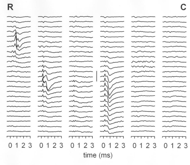
Figure 1. Example of averaged FSPs recorded from 6 electrodes (vertical columns) spaced at 2mm intervals. Traces start at 2.3 mm below the surface and are spaced at 50 µm depths. Vertical scale bar = 20 µV. R – rostral, C – caudal.
Where applicable, experiments conform with Society ethical requirements.