
Introduction
Developing sensory neuronal networks fire spontaneously in preparation to receiving external input (Molnár et al., 2020). Preterm human EEG is characterised by high amplitude activity bursts with discrete voltage distributions across the scalp. Bursts can also be elicited with external stimuli such as touch (Whitehead et al., 2016). While sensory evoked bursts may have voltage distributions similar to those that occur spontaneously, it is not clear whether they involve the same underlying cortical networks. To answer this question, we took advantage of network refractoriness, i.e. that spontaneous activation is followed by a period when the system cannot respond to further input (Fedirchuk et al., 1999).
Methods
We recorded EEG in response to mechanical taps of hands and feet in 35 healthy infants of median 32 weeks corrected gestational age (CGA; range 28-35 weeks) and postnatal age (PNA) 7 days. (CGA = gestational age + PNA). There was a total of 101 stimulation trains (i.e. stimulated limbs) of 2-48 taps (mean 15). We then assessed how magnitude (power spectral density (PSD)) and distribution of endogenous activity preceding the stimulus affected the response to the tap. Ethical approval was obtained from the NHS Research Ethics Committee, and informed written parental consent was obtained prior to each study.
Results
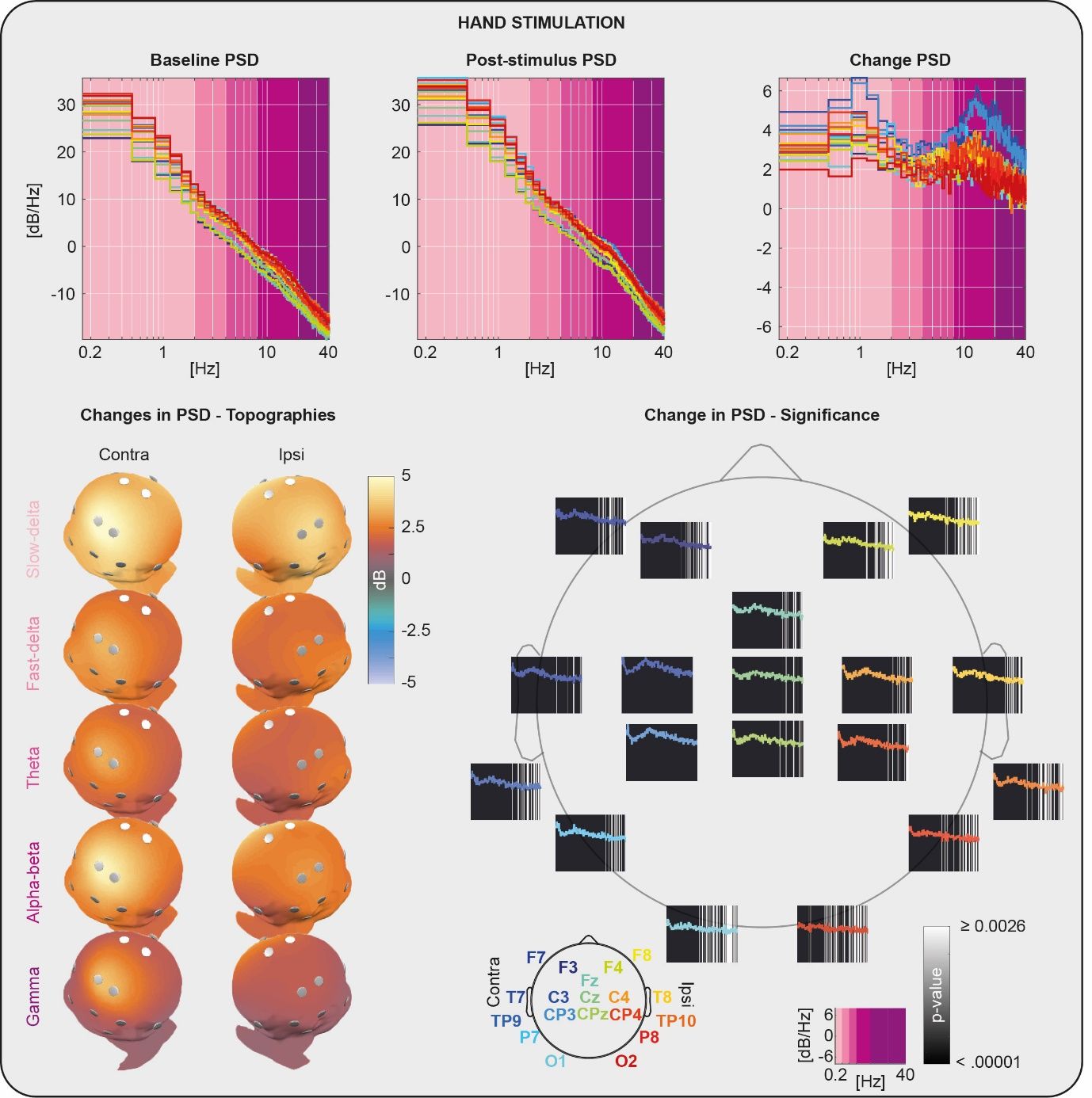
Somatosensory stimulation evoked significant PSD changes in the delta, alpha-beta and gamma bands with peak changes of 6 dB at 1Hz (delta) and 13Hz (alpha-beta). PSD changes at lower frequencies were more widespread across the scalp than those at higher frequencies, which instead were localised at the somatotopic representation of the stimulated limb (i.e. central-contralateral for the hands and central-midline for the feet) (Fig. 1).
The PSD changes evoked in each band by tapping the limbs were inversely related to the PSD present before the stimulus (baseline) and to how similar its distribution was to that of the somatosensory evoked response (SIM) (p < .0001 after correcting for CGA; multiple linear model used: PSD change = b0 + b1 CGA + b2 baseline + b3 SIM + e; adjusted whole model r squared = 0.54 (delta), 0.50 (alpha-beta) and 0.27 (gamma)).
Conclusions
These results indicate that sensory stimulation elicits different oscillatory rhythms, which engage focal or extended networks. These same rhythms are present spontaneously and can make the brain refractory to an incoming external stimulus, which tries to engage the same network. These results are supportive that spontaneous activity in the preterm period represents the activation of maturing sensory cortical architectures in preparation to engagement with the external environment. This offers an etiological explanation for the characteristic discontinuous EEG pattern of preterm neonates.
Future work will examine the implications of these results for the injured preterm brain. Here, spontaneous firing is of higher magnitude than in healthy infants (Whitehead et al., 2016), which correlates with longer subsequent periods of quiescent background, that increases the chance of a poor motor outcome (Koskela, Meek et al. submitted). It is possible that excessive refractoriness could perturb maturation of sensorimotor networks.
Sensory Signals (The Royal College of Physicians, London, UK) (2022) Proc Physiol Soc 50, C19
Poster Communications: Spontaneous cortical activity in somatosensory networks gates their stimulus-driven recruitment in preterm human neonates
Kimberley Whitehead1, Mohammed Rupawala1, Maria Pureza Laudiano-Dray1, Judith Meek2, Sofia Olhede3, Lorenzo Fabrizi1
1University College London, London, United Kingdom 2University College London Hospitals, London, United Kingdom 3Ecole Polytechnique Federale de Lausanne, Lausanne, Switzerland
View other abstracts by:
Where applicable, experiments conform with Society ethical requirements.