Intro: Non-invasive measures of muscle activity are still limited to being used in research environments, although its usefulness in diagnosis and therapeutics is well accepted. One of the factors that might improve its acceptance is ease of use, reliability and reusability without sacrificing ability to capture high-quality data for continuous monitoring.
Aim: We set out to develop a non-toxic, conformal dry electrode using tannic acid, polyvinyl alcohol, and PEDOT:PSS (TPP), as well as a metal-polymer electrode array patch (MEAP) with liquid metal (LM) circuits and TPP electrodes. We then explored the advantageous skin conformability of this array and investigated its potential for recording muscle and tendon location.
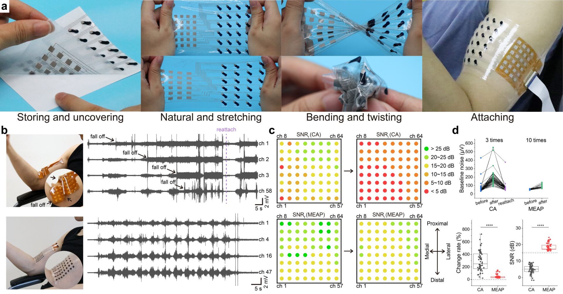
Method: The MEAP fabrication uses screen printers and ovens, ensuring precision and consistency. With use of biocompatible adhesives, the LM circuit were protected within a PDMS casing, with only the TPP electrodes making direct contact with the skin. To test for conformability, we compared our 64 channel MEAP with an identical commercial array (CA) of Ag/AgCl electrodes on a polyimide substrate (Neuracle, China). Data was obtained from five male subjects (23.4 ± 1.9 years). For Ultrasound imaging a Mindray L14-6NE probe (Lysis, Austria) was used.
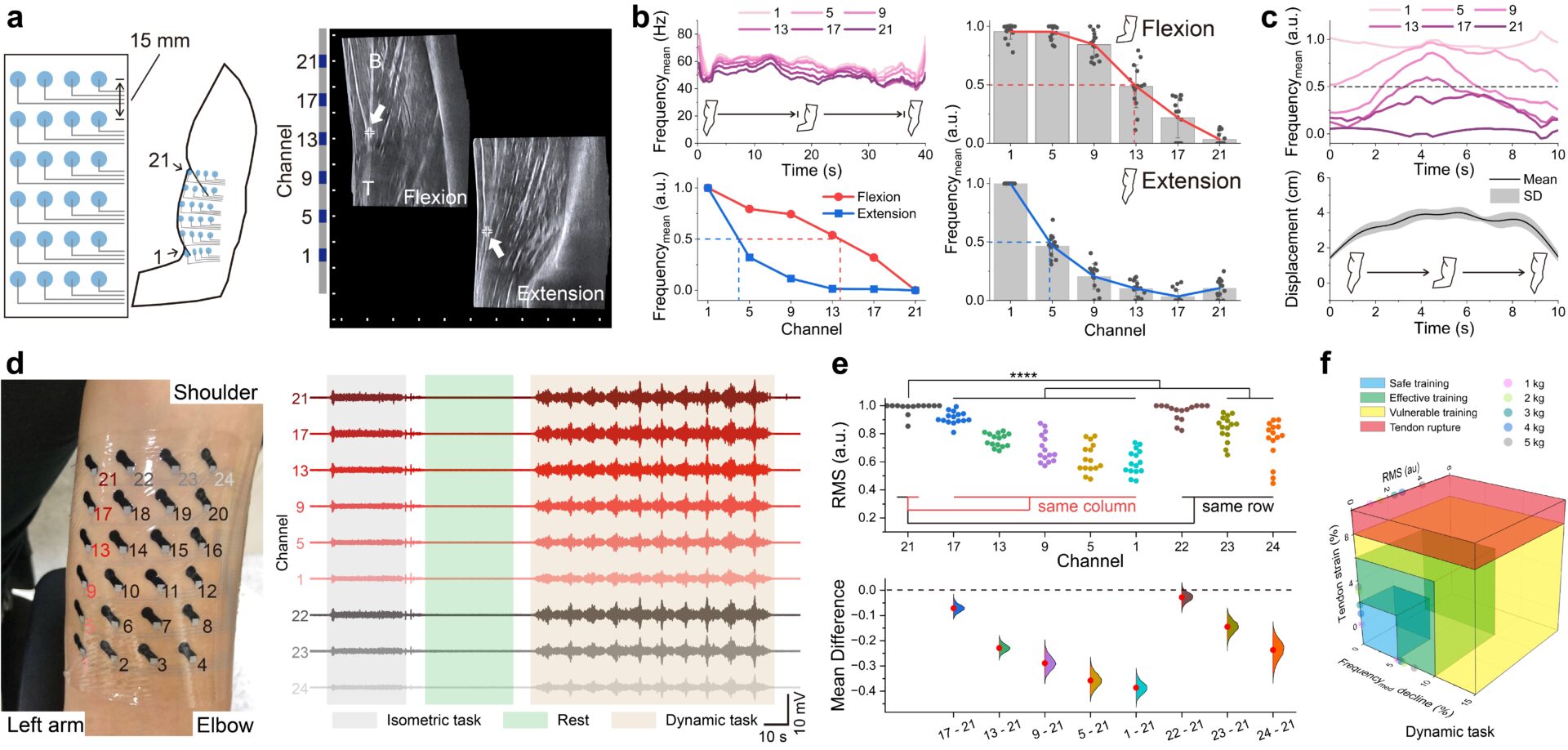
Subjects performed a biceps brachii isometric task, of holding the dumbbell for 30 seconds, with the forearm parallel to the ground forming a right angle with their upper arm. For the dynamic task they performed biceps curl, ranging from full extension to flexion. Subjects were instructed to control the curl speed at approximately 4.5 rad/s for five sessions, with loads ranging from 1 to 5 kg. Tendon strain was calculated as per Walton 2015 (Walton et al 2015) using the distal biceps tendon length. All studies were approved by the Medical Ethics Committee of Southern University of Science and Technology (approval no. 2021SYG049). n=64 for each measurement in Fig.1d; n=15 for Fig.2b, c and e (isometric and dynamic tasks x3, 5 subjects).
Results: Our findings indicate that MEAP exhibits remarkable flexibility, stretchability, and user-friendliness (Fig.1a). When affixed to the muscle-tendon junction, MEAP maintained conformal contact even with skin deformation, contrasting with the noticeable mismatch observed in the case of CA (Fig.1b). This difference in attachment performance was directly reflected in the surface electromyography (sEMG) signals (Fig.1b). MEAP was significantly better than CA for recording sEMG, based on SNR mapping, this is primarily owing to its improved contact even in the presence of substantial skin deformation (Fig1c, d).
Tendon displacement, at the biceps, was quantified using the frequency component of the signal captured by the MEAP, further verified using ultrasound images in an isometric task (Fig.2a, b). Real-time tendon displacements during dynamic tasks were recorded using the MEAPs too (Fig.2c). Each MEAP channel consistently recorded significantly distinct signal in every subject (Fig.2d-f).
Conclusions: These MEAPs, manufactured using scalable screen-printing techniques, feature a completely stretchable material and array architecture, enabling real-time monitoring of muscle stress, fatigue, and tendon displacement. Their potential to reduce muscle and tendon injuries and enhance performance in daily exercise and professional sports holds great promise.