
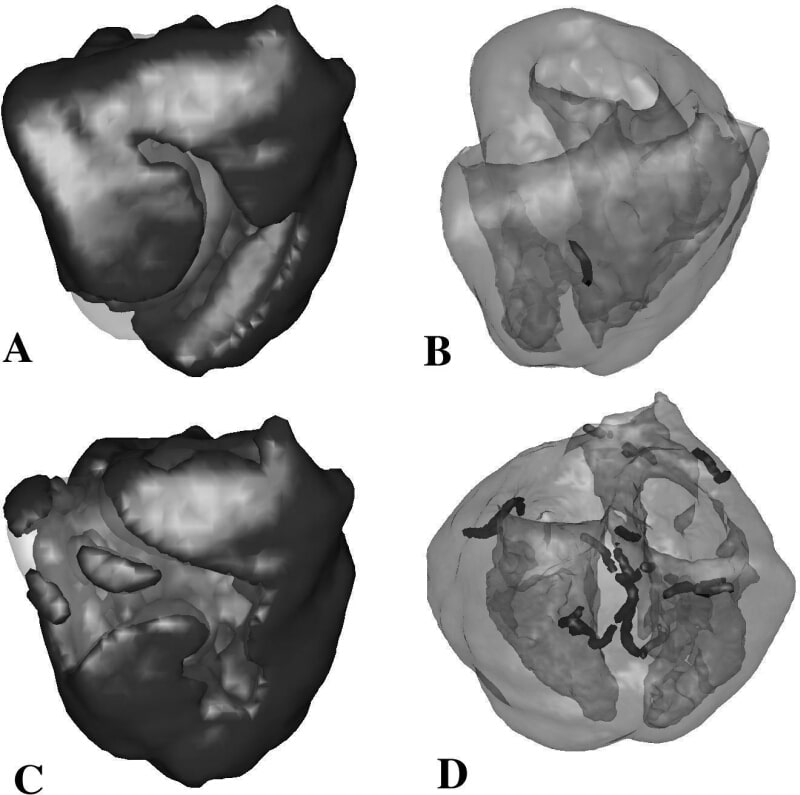
We report on the development of models for the human ventricles. The model is based on anatomical data on the geometry and fibre orientation in the human heart which was gathered and computerized by Hren (1996). It uses the monodomain description of cardiac tissue and employs the ‘weighting functions’ numerical approach, which allows integration of the equations in domains of complex shape with explicit numerical integration schemes. The main framework of the model can be coupled with any type of description of cardiac cells. We discuss different types of such description: low dimensional models, reduced, gamma ionic models and the full ionic models for human ventricular cells. We discuss in details the ten Tusscher at al., (2003) model which is based on recent experimental data on the major ionic currents for human ventricular cells: the fast sodium, L-type calcium, transient outward, rapid and slow delayed rectifier, and inward rectifier current. The model includes intracellular sodium, potassium and calcium dynamics, allowing for the realistic modelling of calcium transients, frequency dependence of the intracellular sodium concentration, and the positive contraction staircase typical for human ventricular myocardium. Using this model we study normal and abnormal wave propagation. We show that for normal parameter values the re-entrant arrhythmia (scroll wave) persists in the course of time and does not break-up into fibrillation. The dynamics of the scroll wave is meandering, however the meandering of the scroll wave rotating in the right ventricle is less pronounced than that in the left ventricle of the heart. The computed electrocardiogram (ECG) in the right ventricle is similar to that of monomorphic tachycardia, while the ECG of a scroll wave rotating in the left ventricle is similar to that of polymorphic tachycardia. The upper row of the figure shows the scroll wave pattern of excitation in the right ventricle of the heart (A) and the corresponding filament (B). By modifying the properties of the cardiac tissue, we were able to find parameters for which the scroll wave becomes unstable and breaks down into a multiple wavelet pattern that is considered to be a model of ventricular fibrillation. We compare the results of these simulations with the recent clinical data by Nanthakumar et al.(2004), who performed epicardial recordings of excitation patterns during fibrillation in the human heart using a 4x5cm electrode plaque. First, we show that the ECG pattern occurring in our model is similar to that during ventricular fibrillation and that its dominant frequency is close to that recorded by Nanthakumar et al. (2004) and others. We also compare the wavelet dynamics occurring in the 4x5cm left ventricular epicardial patch in our model to those observed by Nantakumar et al. We find that surface re-entry can be observed during approximately 10% of the time, and find large activation fronts following repeatable pathways, smaller fractionated wave fronts and epicardial breakthrough waves, similar to the observations by Nanthakumar et al. Finally, we study the role of re-entry in the three-dimensional organization of fibrillation in the ventricles by counting the number of filaments, the organizing centers of scroll waves and hence a measure for the number of re-entrant sources present. We determine the number of filaments as a function of time, compute their individual and total lengths and compare them with previous simulations using a canine ventricular geometry. The bottom row of the figure shows a typical activation pattern during fibrillation (C) and the scroll wave filaments present at that same moment (D). Our study supports the results by Nanthakumar et al. (2004) that ventricular fibrillation in the human heart may be organized by only a small number of sources.
University of Oxford (2004) J Physiol 561P, SA15
Research Symposium: STUDY OF RE-ENTRANT ARRHYTHMIAS IN ANATOMICALLY BASED MODELS OF THE HUMAN HEART
Panfilov ,Sasha ; ten Tusscher,KHWJ ;
1. Division of Mathematics, University of Dundee, Dundee, United Kingdom. 2. Department of Theoretical Biology, Utrecht University, Utrecht, Netherlands.
View other abstracts by:
Where applicable, experiments conform with Society ethical requirements.