In congestive heart failure (CHF), ventricular arrhythmias are often caused by triggered activity (Tomaselli & Marban, 1999). Nuss et al. (1999) described spontaneous depolarizations (SD) that occur during electrical diastole in single ventricular myocytes isolated from a canine CHF model and hypothesized that a chronically activated depolarizing current may cause them. A volume-sensitive, inwardly rectifying, poorly selective cation current (ICir,swell) was identified in the same canine model (Clemo et al. 1998). ICir,swell is a candidate for the current underlying SD because it is chronically activated under isosmotic conditions in the setting of CHF.
To test this hypothesis, the correlation between ICir,swell activity and frequency of SD was examined in single ventricular myocytes obtained from a rabbit aortic regurgitation model of CHF and from normal rabbits. The amphotericin perforated-patch clamp technique was used to avoid unpredictable cell swelling and changes in membrane currents that often slowly occur with ruptured patches. Ramp voltage clamps (28 V s-1) were applied to determine current-voltage (I-V) curves. Current clamp (2-5 nA, 2 ms pulse) was used to elicit a train of action potentials (AP) at 0.2 Hz for 50 s, and then the stimulus was turned off to allow detection of SD (> 5 mV depolarization). The activity of ICir,swell was decreased by osmotic manipulation of cell volume or by addition of the ICir,swell blockers Gd3+ (Clemo & Baumgarten, 1997) or GsMTx-4, a G. spatulata peptide toxin (Suchyna et al. 2000).
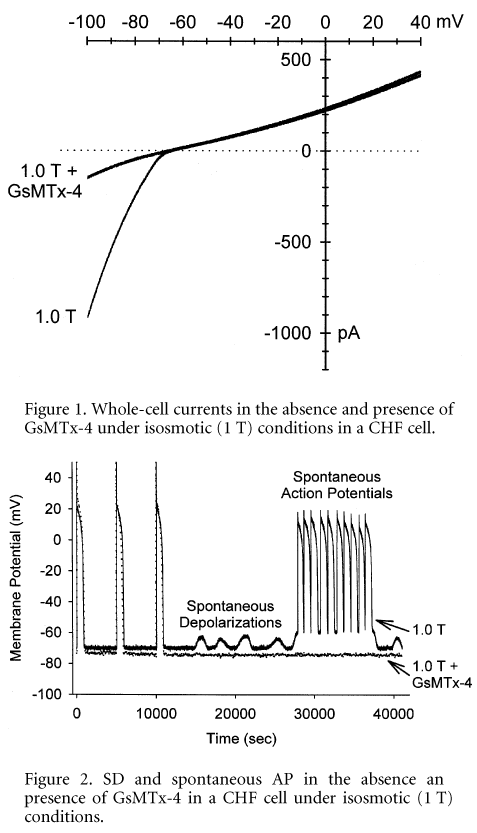
Under isosmotic conditions (1.0 T), an inwardly rectifying current that was attenuated by hyperosmotic (1.5 T) cell shrinkage was seen in 25 of 30 CHF cells. The difference current at -80 mV was -2.9 ± 0.10 pA pF-1. Application of GsMTx-4 (0.4 mM, graciously provided by Dr Fred Sachs, SUNY Buffalo, USA) significantly reduced this inward rectifying current, as shown in Fig. 1. The GsMTx-4-sensitive current at -80 mV was -1.8 ± 0.09 pA pF-1 (n = 10). Similar results were found for Gd3+ (10 mM). Hyperosmotic shrinkage and application of ICir,swell blockers had no effect on whole-cell currents of normal cells.
Following a train of stimulated AP, SD were seen in 24 and spontaneous AP in 15 of the 30 CHF cells, as demonstrated in Fig. 2. In contrast, a train of AP elicited SD in only 2 and spontaneous AP in 0 of 30 CHF cells after ICir,swell was blocked by shrinkage in 1.5 T. Moreover, in cells with SD, the ICir,swell blockers Gd3+ and GsMTx-4 reduced the occurrence of SD by 85 and 90 %, respectively. In normal myocytes under isosmotic conditions, neither SD nor spontaneous AP were observed.
This study showed that: (1) ICir,swell is chronically active under isosmotic conditions in ventricular myocytes taken from a rabbit aortic regurgitation model of CHF; (2) under the same conditions, SD and spontaneous AP are present; and (3) attenuation of ICir,swell reduces the frequency of SD and spontaneous AP. These results suggest that chronic activation of ICir,swell may underlie the genesis of SD in failing ventricular myocytes and may potentiate triggered cardiac arrhythmias in the setting of heart failure. Novel blockers of ICir,swell such as GsMTx-4, which inhibits atrial fibrillation in an isolated, perfused rabbit heart model (Bode et al. 2001), also may be efficacious in preventing the triggered arrhythmias seen in CHF.