
High-resolution three-dimensional (3-D) images of myocytes and collagen have been acquired across the left ventricular (LV) wall in seven normal rat hearts using extended-volume confocal microscopy1. One adult male Wistar and six Wistar-Kyoto (WKY) rats were anaesthetised with 5% halothane and the hearts exposed through a thoracotomy. Hearts were heparinised, excised and perfusion-stained with picrosirius red dye (0.1% Sirius Red F3BA in picric acid). After 5-7 days fixation in Bouin’s solution, a small transmural block was cut from the lateral LV free wall, dehydrated in graded ethanols, and embedded in agar resin2. The embedded tissue block was mounted on a high-precision three-axis stage and imaged using confocal microscopy at 0.4-1.22 µm resolution to a depth of 35 µm. The surface was then milled to remove 30 µm and the process repeated. Overlapping image volumes were processed and assembled to give 3-D image volumes up to 4 mm x 1 mm x 1 mm. Collagen was segmented from each volume using techniques which identify voxels that are bright relative to the local background. Volumetric analyses of collagen density and arrangement were performed, together with comparisons on curved transmural cutting planes that present consistent views relative to the varying fibre orientation. Analysis of fibre orientation and collagen fraction showed no difference between Wistar and WKY hearts. Results are presented as mean±S.E.M., n=7. 1 factor ANOVA showed no significance for collagen fractions sampled from five wall depths (7, 25, 50, 75, 93%). Images of tissue and collagen in the Wistar heart at 7% (sub-epicardial), 50% (midwall), and 75% (sub-endocardial) locations are shown in Figure 1. Cross-sectional collagen alignment was computed in these three regions. A Student’s t-Test showed no significant difference (p=0.44) in the normalised variance of alignment between the midwall and sub-endocardial regions (0.032±0.006), but a significant difference (p=0.001) between those and the normalised variance of alignment in the sub-epicardial region (0.014±0.002). The high degree of alignment corresponded with the presence of collagen lining the surfaces of cleavage planes between laminae in the midwall and sub-endocardium, and low alignment with isolated collagen strands in the sub-epicardium where laminae were not present. The percentage of the wall exhibiting this lack of laminae (and corresponding decreased alignment) ranged between 10% and 25%. Analysis of 3-D images has demonstrated that while perimysial collagen fraction is approximately the same across the wall, the form varies transmurally in correspondence with the underlying laminar architecture.
University of Cambridge (2008) Proc Physiol Soc 11, PC68
Poster Communications: Transmural organisation of perimysial collagen in the rat left ventricle
G. Sands1, A. Pope2, B. Smaill1,2, I. Legrice1,2
1. Bioengineering Institute, University of Auckland, Auckland, New Zealand. 2. Department of Physiology, University of Auckland, Auckland, New Zealand.
View other abstracts by:
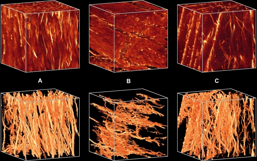
Figure 1. Sub-epicardial (A) midwall (B) and sub-endocardial (C) blocks showing myocytes and collagen (top) and collagen only (bottom). Block dimensions are 300 µm at 1.22 µm resolution.
Where applicable, experiments conform with Society ethical requirements.