
It has been suggested that trimethylamine oxide (TMAO), a liver oxygenation product of gut bacteria-produced trimethylamine (TMA), may be a marker and mediator of cardiovascular diseases. However, the mechanisms of plasma TMAO increase and its biological effects are obscure. We evaluated the effect of age, a cardiovascular risk factor, on rat plasma levels of TMA and TMAO, and cytotoxicity of the two molecules in human vascular smooth muscle cells (VSMCs). The experiments were performed on (i) 3-month-old Sprague Dawley rats (n=8) and Wistar-Kyoto rats (n=8) and (ii) 18-month-old, male, Sprague Dawley rats (n=8) and Wistar-Kyoto rats (n=8), maintained on the same standard laboratory diet and in the same environmental conditions. The rats were anaesthetized i.p. with urethane 1.5 kg/bw (Sigma-Aldrich) for a direct arterial blood pressure and intestinal blood flow measurements. Next, blood samples were taken from the right ventricle of the heart and rats were killed by decapitation. A segment of the colon (7-8 cm, a middle part between the cecum and the rectum) was harvested for histological evaluation. The colon content (stool masses) was collected for biochemical and microbiome analysis. Gut bacteria were analyzed with a 16S ribosomal ribonucleic acid gene sequence analysis. TMA and TMAO were measured in the colon content, portal blood and systemic blood using chromatography coupled with mass spectrometry. Viability of VSMCs exposed to TMA and TMAO at concentrations found in rats, and higher, was evaluated with MTT test. Older rats showed significantly different gut bacteria composition (Fig. 1) and higher, but not significantly, TMA stool level than younger rats. Systemic blood TMA level was significantly higher in older rats, which was associated with a significantly higher gut-to-blood TMA penetration (a significantly higher ratio of portal blood to stool concentration of TMA). There was no significant difference in arterial blood pressure between the groups. In contrast, older rats showed significantly lower intestinal blood flow and significantly reduced thickness of colonic mucosa which constitutes the physical barrier for gut-to-blood penetration of bacterial products. Older rats showed lower, but not significantly lower, TMA to TMAO liver oxidation and plasma TMAO level. In vitro, TMA at concentration of 500 µmol/L (2-fold higher than in portal blood) significantly decreased VSMCs viability (Fig. 2). In contrast, TMAO at 1000-fold higher concentration than those observed under physiological conditions in rat plasma, had no effect on VSMCs viability. In conclusion, older rats show higher plasma level of TMA due to increased gut-to-blood penetration of the bacterial metabolite. TMA, but not TMAO, affects VSMCs viability at concentrations close to physiological ones. Therefore, we would hypothesize that it is TMA, not TMAO, that exerts detrimental effects on cardiovascular system and may contribute to increase in cardiovascular risk with aging.
Physiology 2019 (Aberdeen, UK) (2019) Proc Physiol Soc 43, PC245
Oral Communications: Trimethylamine, a gut bacteria metabolite, increases in rat plasma with age and affects vascular smooth muscle cells viability.
K. Jaworska1, M. Konop1, T. Hutch1, K. Perlejewski3, M. Grochowska3, M. Radkowski3, A. Bielak-Zmijewska2, G. Mosieniak2, E. Sikora2, M. Ufnal1
1. Department of Experimental Physiology and Pathophysiology, Medical University of Warsaw, Warsaw, Poland. 2. Laboratory of Molecular Bases of Aging, Nencki Institute of Experimental Biology, Polish Academy of Sciences, Warsaw, Poland, Warsaw, Poland. 3. Department of Immunopathology of Infectious and Parasitic Diseases, Medical University of Warsaw, Warsaw, Poland.
View other abstracts by:
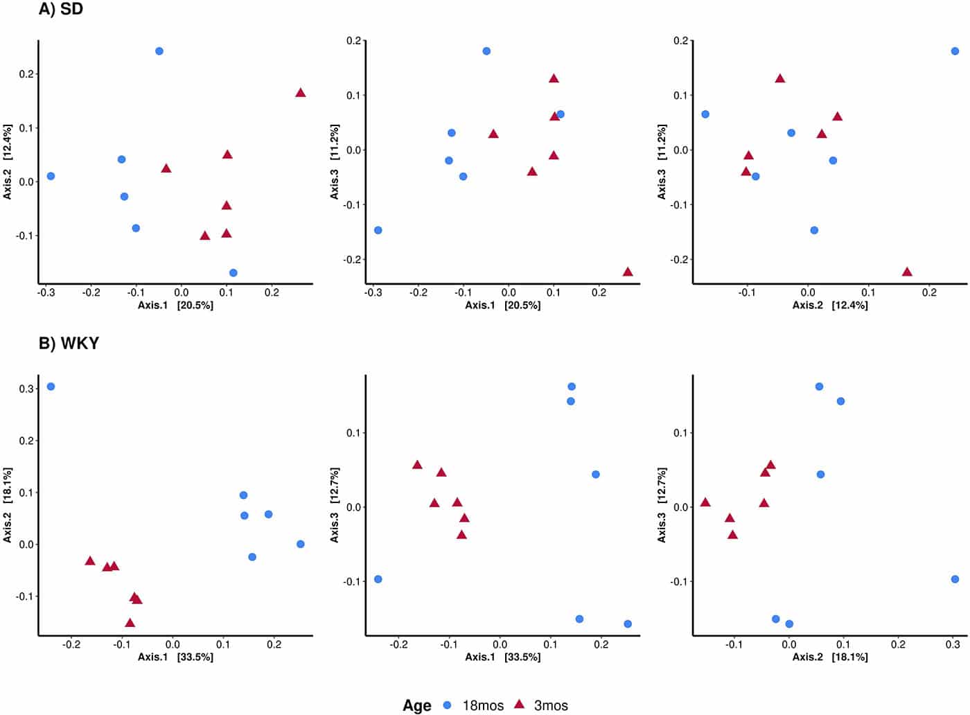
Principal Coordinate Analysis (PCoA) clustering according to similarity of gut microbiota composition in stool samples. Percentage value (%) on each axis indicates % of the captured variation in the input data. PCoA analysis indicates that samples ordinated closer to one another are more similar than those ordinated further away. PCoA shows how examined samples differ between each other in tri-dimensional space, forming distinct clusters.
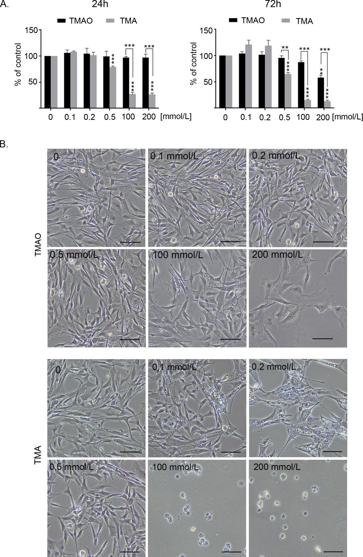
Fig. 2. A. Comparison of viability of human vascular smooth muscle cells (hVSMCs) treated with trimethylamine oxide (TMAO) or trimethylamine (TMA). MTT test was performed after 24 and 72h of treatment. Results were normalized to the control (untreated) cells; Graphs show mean �SE, ** p< 0.01, ***p<0.001; B. Representative pictures showing changes in the number and morphology of cells after 72 hours of treatment with of trimethylamine oxide (TMAO) or trimethylamine (TMA), bar 100�m.
Where applicable, experiments conform with Society ethical requirements.