
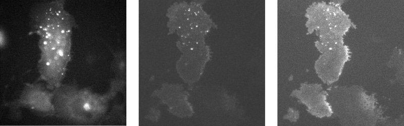
Many facets of cell physiology are now accessible for study by live cell imaging. To a large extent this is attributable to the growth in availability of cellular and sub-cellular fluorescent probes. These probes take many different forms; they may localize to specific environments within a cell, they may detect changes in ion concentration or they may be used to tag specific proteins, thereby enabling tracking and localization of the tagged protein. Whilst fluorescence detection technology has also developed rapidly in recent years, one major limitation of conventional imaging remains the lack of depth resolution. The comparatively new technique of total internal reflection fluorescence (TIRF) microscopy overcomes this limitation, using the principle of total internal reflection at a glass/water interface to generate an evanescent wave and excite a very thin fluorescent volume immediately above the interface (Axelrod, 2003). The illumination depth using this method is extremely small, allowing fluorescence excitation up to only a few hundred nanometres into the sample, typically a small fraction of the thickness of a cell. Small changes to the angle of incidence of the incoming light beam to the interface alter the penetration depth of the evanescent wave, and this effect can be utilised to probe changes in fluorescent signal pattern at a range of depths within the TIRF interface. In this study we devised a new TIRF system that allows rapid changes in the depth of measurement into the cell. This enables distinctions to be made between fluorescence arising due to changes at the cell membrane and those arising from changes within the cell. The rapid depth changes are achieved by use of a galvanometer mounted mirror deflecting the beam at a variable angle into a prism below the cell via a combination of lenses. Depth changes can be achieved on a millisecond timescale allowing a dynamic event to be tracked almost simultaneously at different depths. The images shown are those of COS cells transfected with YFP-e-sarco-glycan. The epifluorescence image (panel 1) shows general cell wide labelling whereas the TIRF images (panels 2 and 3 taken with calculated illumination depth constants of 62 and 80nm), show that the protein is differentially expressed within the cell. Images were acquired sequentially with 50ms intervals between frame starts. These results illustrate that variable angle TIRF microscopy can be used to collect multiple thin optical sections close to the cell membrane surface. This has the considerable advantage over conventional TIRF microscopy that dynamic changes, such as the movement of structures towards or away from the cell membrane, can be discriminated in near real time.
University of Bristol (2005) J Physiol 567P, PC186
Poster Communications: Variable Depth Total Internal Reflectance Fluorescence (TIRF) Microscopy in COS Cells
Reynolds, Martyn A; Cavazzini, Michele; Emptage, Nigel; Thomas, Martin V;
1. Research & Development, Cairn Research Ltd, Faversham, Kent, United Kingdom. 2. Pharmacolgy, University Of Oxford, Oxford, United Kingdom.
View other abstracts by:
Where applicable, experiments conform with Society ethical requirements.