
In native tissues, relatively low protein expression levels and overlapping sensitivities of antibodies within families of GPCRs present challenges to transfer of knowledge of receptor mechanisms from cell culture. In contrast, in recombinant systems epitope tags and fluorescent proteins provide powerful and sensitive means of tracking the concentration, location and movement of receptors. Two approaches have now started to close this gap. First is the use of fluorescent ligands, which can be tested and validated in recombinant systems [1] and then employed in intact, live native tissue using confocal microscopy [2-4]. Agonist and antagonist ligands can be employed, each conferring advantages and disadvantages. The second approach is the creation of mouse strains expressing transgenic receptors labeled with fluorescent proteins [5]. Examples will be given illustrating the current state of play with these approaches on alpha-1, alpha-2 and beta-adrenoceptors, and angiotensin receptors, mainly using rat and mouse blood vessels, in which fluorescent ligand binding shows that a range of cell types express adrenoceptors, e.g. nerves, adventitial fibroblasts, smooth muscle, endothelial and fat cells. The distribution of receptors is shown to be both in intracellular organelles and the cell surface, the consequences of dimerisation can be explored in receptor KOs and phenomena such as binding kinetics and receptor internalization and trafficking can be followed in real time in native tissues. These approaches are steadily closing the gap between cell culture and native tissues. In the particular example of blood vessels, these approaches have shown that receptors exist on types of cells where they were previously not known to exist. Receptors for vasoactive agents such as catecholamines and angiotensin II have been found on adventitial and endothelial cells as well as where they were expected, on smooth muscle. This has led to new avenues of research into vascular control. An example is shown in figure 1, where adrenoceptors are shown on the endothelial cells of mouse carotid artery as well as on smooth muscle.
Life Sciences 2007 (2007) Proc Life Sciences, SA76
Research Symposium: Visualisation of receptors in native tissue
I. McGrath1, J. D. Pediani1, L. Methven1, J. MacMillan1, C. J. Daly1
1. Autonomic Physiology Unit, University of Glasgow, Glasgow, United Kingdom.
View other abstracts by:
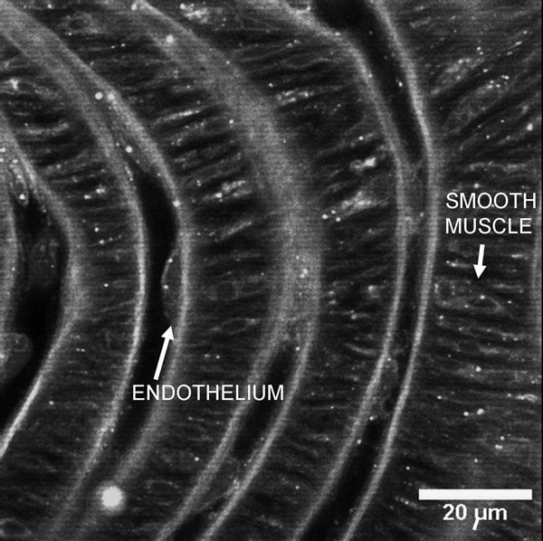
A confocal 'slice' through the ridged luminal surface of a mouse carotid artery. The alternating vertical 'bands' show the repeating sequence of (from left) luminal space endothelium internal elastic lamina and medial smooth muscle. The continuous curving vertical white lines show the natural fluorescence of the elastic lamina. All other staining indicates the binding of a fluorescent ligand to α1-adrenoceptors which can be seen on both endothelial and smooth muscle cells.
Where applicable, experiments conform with Society ethical requirements.