
Physiology News Magazine

Being an artist and a scientist
News and Views
Being an artist and a scientist
News and Views
Stefanie Reichelt
University of Cambridge, UK
https://static.physoc.org/app/uploads/2020/01/28104950/pn.95.16.figure1.jpg
I am a Cambridge-based researcher and photographer. I have always been an observer and a dreamer, and I have combined the qualities of both to become a scientist and a photographer.
For me art and science have never been separate but complimentary ways of seeing. As a biology undergraduate, I learned the craft of scientific illustration, producing accurate but beautiful drawings and paintings of beetles and butterflies. Photography was a means to document processes in Petri dishes as well as in life. Electron microscopy and light microscopy both use large format film, and dark room work was essential. I experimented with a pinhole camera and making my own photographic paper processes.
For 5 years I worked with one of the UK-based inventors of the confocal microscope, Brad Amos, at the MRC-LMB in Cambridge. As a biologist I had not been trained to take microscopes apart and put them back together, but soon became caught up in optical design and testing work. I was one of the first to use a 405 nm (violet) laser in a confocal microscope and also worked on a high-resolution spectral confocal development, as well as a compact point-scanning confocal microscope, which was commercialized by a company (BioRAD).
Since 2005 I have been the Head of the Light Microscopy Laboratory at the CRUK Cambridge Research Institute. My research includes the development of new imaging techniques, which will enable the visualisation of molecules in cells for cancer diagnostics. See www.cruk.cam.ac.uk/core-facilities/light-microscopy-core
My group at the CRUK Cambridge Institute uses confocal point-scanning, spinning disc systems and non-linear imaging techniques to study live cell divisions and cell development, tumour development and biopsy material. As head of the Microscopy Laboratory, I am in charge of a team of imaging specialists who work in collaboration with researchers, obtaining results that are crucial to developing new treatments for cancer patients.
Last year we developed with a group of physicists a CARS Raman multiphoton imaging system, which could help in eliminating the bane of staining in translational imaging experiments.
For over 300 years optical microscopy has been one of the fundamental tools to study biology. Staining with dyes or fluorescent labels has remained one of the main ways of visualisation and diagnostic imaging of cells ever since the first use of dyes in the mid-nineteenth century. However, many such stains affect the normal function of cells making unfeasible their use with live cells.
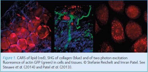
The multi-photon microscopy set-up at the CRUK Cambridge Institute is helping researchers to image cell and tissue structures in live cells without the requirement of stains. Through the overlaying of lasers and exploiting how tissues respond to certain wavelengths, we can acquire structures such as collagen, muscle and lipids stain free (Fig. 1). These processes are acquired through techniques called second harmonic generation (SHG) and coherent anti-stoke raman scattering (CARS).
In a similar concept to the function of a microwave oven, where absorption of microwaves causes vibration of water molecules and heating of food, CARS works by the absorption of light and detection of vibrational movements of molecules. This opens up a whole field of potential stain free imaging, currently unavailable by conventional techniques. Imaging drug localisation is capable of furthering understanding of pharmacodynamic properties in treating diseases.
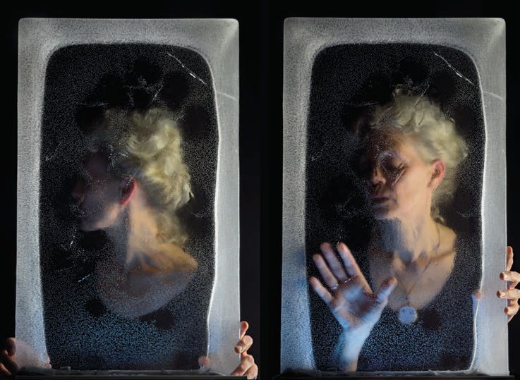
My photographic work explores perception in photography and our conscious and unconscious reactions to the images we see. It raises such questions as: is seeing believing? is what we see what we perceive? and what is the relationship between our subjective and objective realities? Leaving photographic interpretations to observers’ imaginations, as with fairy tales, I seek to depict photographic stories with clues, hints and surprises, so that the perception of what lies within the photographs elicits desires, fears and unease in the observer. I have exhibited widely and my projects are features in photography and science magazines.
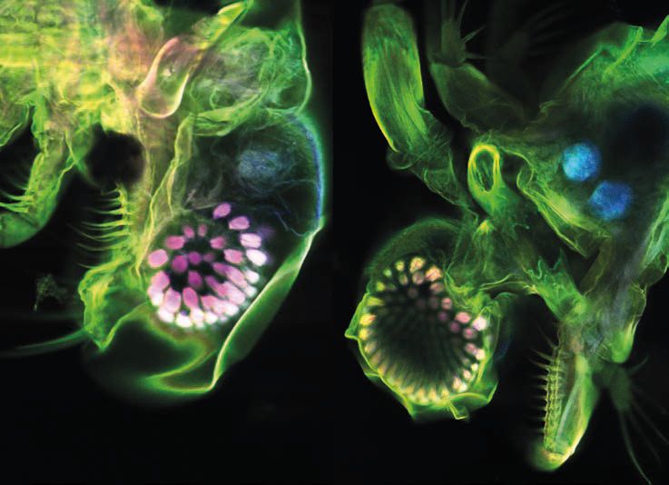
As a scientist I take microscopic images of the human body, of small cells that move and divide, and contain even smaller molecules. These objective images, both informative and beautiful, enhance the knowledge of our physical self and help researchers develop cures for cancer. My images were projected onto Senate House during the 800-year Cambridge Anniversary Celebration and are part of the science photo library collection
(vimeo.com/8768073).
I make hand-made photo books from my projects and also experiment with alternative display options using LCD screens or viewing boxes with sound (www.stefaniereichelt-photographyandprints.com/projects.php) (Fig. 2 & 3). A recent project ‘Traces of Genius’ can be found here: tracesofgenius.wordpress. com. The aim was to record the ‘traces’ left behind by the scientists of the famous MRC Laboratory of Molecular Biology and create a visual ‘memento mori’ for the old LMB building. The project has resulted in a photographic archive of the human traces and marks in the vacated old LMB building. These were exhibited in the new LMB building following the scientists’ move from the old to the new space. A collaborative literary art book will be published soon combining photographs from the project with short stories inspired by them.
When joining CRUK, I founded ArtCell Gallery, which provides exhibition space on the Addenbrooke’s Hospital site for local artists and the science community. I am the curator and have organized and designed the ArtCell exhibitions and events (www.stefaniereichelt -photographyandprints.com/artcell.html).
In March 2014, as part of the 2014 Cambridge University Science Festival, I organized and hosted a day of talks, lectures, demonstrations and exhibitions on ‘the Art of Scientific Imaging’ with the Royal Photographic Society. As part of the festival the ‘International Images for Science exhibition 2013’ will be on display until May. We also had the amazing Camper Obscura to demonstrate the basic principles of imaging: camperobscura.co.uk
References
Patel II, Steuwe C, Reichelt S & Mahajan S (2013). Coherent anti-Stokes Raman scattering for label-free biomedical imaging. J Opt 15, 094006.
Steuwe C, Patel II, Ul-Hasan M, Schreiner A, Boren J, Brindle KM, Reichelt S & Mahajan S (2014). CARS based label-free assay for assessment of drugs by monitoring lipid droplets in tumour cells. J Biophotonics (in press, doi: 10.1002/jbio.201300110).