
Physiology News Magazine
Endocrine granules
Exocytosis and endocytosis are not as separate as once thought, report David Perrais and colleagues
Features
Endocrine granules
Exocytosis and endocytosis are not as separate as once thought, report David Perrais and colleagues
Features
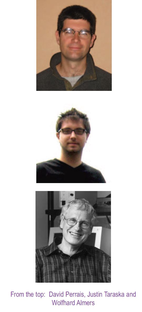
David Perrais (1), Justin Taraska (2), & Wolfhard Almers (3)
1: Cellular Physiology of the Synapse, CNRS and Université Bordeaux 2, France
2: Department of Physiology and Biophysics, University of Washington, Seattle, Washington, USA
3: Vollum Institute, Oregon Health and Science University, Portland, Oregon, USA
https://doi.org/10.36866/pn.58.33
Endocrine cells release proteins and small molecules into the blood stream. It is well known that the release is controlled by regulating exocytosis, the fusion of secretory granules to the plasma membrane. As in presynaptic terminals, exocytosis is triggered by an increase in cytoplasmic Ca2+. The classical view was that, once exocytosis has occurred, the membrane of the vesicle flattens into the plasma membrane and mixes with it, and that the material thus inserted into the plasma membrane is later retrieved by endocytosis. This view began to change when capacitance measurements could detect the exocytosis of single granules and, more recently, when the method was combined with amperometry to track exocytosis and catecholamine release simultaneously (Ales et al. 1999). At least occasionally, individual granules were seen to connect transiently with the plasma membrane, release their catecholamine and then reseal. This transient exocytosis is sometimes called ‘kiss and run’ (Ales et al. 1999) or ‘cavicapture’, because the cavity of the granules is recaptured intact (Taraska & Almers, 2004). In small synaptic terminals that have only about a hundred synaptic vesicles, the benefits of kiss-and-run seem obvious, as this mechanism would allow cells to re-use synaptic vesicles rather than making new ones. Endocrine granules, however, are not generally believed to be re-used. Nor does cavicapture seem to influence how much catecholamine is released by granules. So why do endocrine cells perform cavicapture, and what difference does it make physiologically?
Endocrine cells also release peptides and proteins. When these are fused to green fluorescent protein (GFP), their release from single granules can be observed by evanescent field fluorescence microscopy (also called total internal reflection fluorescence, or TIRF). The evanescent field illuminates only a thin surface layer where the cell comes in close contact with the glass coverslip, and leaves deeper regions in the dark. Our group has used it to compare the release of two proteins from single granules, neuropeptide Y and the protease tissue plasminogen activator, or tPA, both normally contained in chromaffin granules (Perrais et al. 2004). NPY-GFP was lost from granules in fractions of a second while tPA-GFP remained there. About one-third of the granules re-sealed before they could release all their tPA-GFP.
Related findings were made in cell lines derived from chromaffin cells (Taraska et al. 2003) and pancreatic β cells (Tsuboi et al. 2004). Thus, cavicapture keeps some proteins in granules after catecholamines, and in most cases NPY, are released completely. Surprisingly, some granules were seen to undergo exocytosis, then cavicapture, and then to release the remainder of their tPA in a second round of exocytosis. Related results were also obtained in PC12 cells (Holroyd et al. 2002). Apparently, granules are sometimes re-used after all. And perhaps cells regulate protein release not only by controlling exocytosis, but also by cavicapture. Other recent studies highlight further complexities of protein release from single granules (e.g. Barg et al. 2002).
Do granules get to keep their membrane components when they undergo cavicapture? They probably exchange most or all of their lipids with the plasma membrane (Taraska & Almers, 2004), but keep selected membrane proteins such as phogrin. Most granules also lose the majority of their synaptobrevin-2 and synaptotagmin-1 (Tsuboi et al. 2004), although one supposes that granules planning to undergo exocytosis a second time must retain enough of both.
Finally, which proteins are required for cavicapture? Some proteins participating in clathrin-mediated endocytosis, among them clathrin itself, never appear at sites of cavicapture, but dynamin does. Dynamin separates endocytic vesicles from the plasma membrane. When dynamin is blocked, then cavicapture is blocked as well, and phogrin and tPA are both rapidly lost from granules (Holroyd et al. 2002, Tsuboi et al. 2004).

Cavicapture implies that exocytosis and endocytosis are not as clearly separated as they are, e.g. in the recycling of transferrin receptors between endosomes and plasma membrane. Instead, there are multiple steps on a continuum (Fig. 1). When an exocytic fusion pore connects the inside of the vesicle to the outside of the cell, catecholamines are released within 100 ms, as judged by amperometry. However, the open cavity of many granules stays behind (Taraska et al. 2003), and may re-seal in seconds to minutes (Perrais et al. 2004). A fission machinery including dynamin is recruited. Such recruitment is probably more important for cavicapture than the size of the fusion pore, because during its life the pore can start out small enough to retard the escape of catecholamines, then dilate to allow partial escape of a relatively large protein such as tPA-GFP, and still close again.
It remains to be seen what mechanisms control how soon the pore closes, and how large it gets before it closes. Dysfunctions in such control mechanisms could alter the release of proteins and hormones from endocrine cells, and possibly lead to abnormal protein release.
References
Ales E, Tabares L, Poyato JM, Valero V, Lindau M & Alvarez de Toledo G(1999). High calcium concentrations shift the mode of exocytosis to the kiss-and-run mechanism. Nat Cell Biol 1, 40-44.
Barg S, Olofsson CS, Schriever-Abeln J, Wendt A, Gebre-Medhin S, Renstrom E & Rorsman P (2002). Delay between fusion pore opening and peptide release from large dense-core vesicles in neuroendocrine cells. Neuron 33, 287-299.
Holroyd P, Lang T, Wenzel D, De Camilli P & Jahn R (2002). Imaging direct, dynamin-dependent recapture of fusing secretory granules on plasma membrane lawns from PC12 cells. Proc Natl Acad Sci USA 99, 16806-16811.
Perrais D, Kleppe IC, Taraska JW & Almers W(2004). Recapture after exocytosis causes differential retention of protein in granules of bovine chromaffin cells. J Physiol 560, 413-428.
Taraska JW & Almers W (2004). Bilayers merge even when exocytosis is transient. Proc Natl Acad Sci USA 101, 8780-8785.
Taraska JW, Perrais D, Ohara-Imaizumi M, Nagamatsu S & Almers W (2003). Secretory granules are recaptured largely intact after stimulated exocytosis in cultured endocrine cells. Proc Natl Acad Sci USA 100, 2070-2075.
Tsuboi T, McMahon HT & Rutter GA (2004). Mechanisms of dense core vesicle recapture following ‘kiss and run’ (‘cavicapture’) exocytosis in insulin-secreting cells. J Biol Chem 279, 47115-47124.