
Physiology News Magazine

Fluorescence imaging systems: A quick overview of the technology.
Features
Fluorescence imaging systems: A quick overview of the technology.
Features
John Dempster & David Wokosin
Centre for Biophotonics, University of Strathclyde
https://doi.org/10.36866/pn.48.12


In the past decade, the ability to visualise specific probes within a biological specimen has brought about a Renaissance of light microscopy. Considerable developments in staining techniques now allow a high degree of specificity by labelling, and subsequently imaging. Currently most physiological applications arise in the study of intracellular calcium dynamics using the fluo dyes and also intracellular structures tagged with green fluorescent protein. The newcomer to imaging is presented with an array of systems, ranging from relatively simple cameras, through various types of confocal microscope, to the two photon excitation (2P) microscope, not to mention more specialised techniques such as total internal reflection (TIRF) or fluorescence resonance energy transfer (FRET). Not surprisingly, it can be difficult to determine the relative merits of different offerings, or separate marketing hype from fact. Given the painfully high cost of some systems, making the right choice is no small matter.


The three most important issues concerning the choice of system are – depth, speed and invasiveness. Events of interest don’t always lie on the surface of the specimen, requiring the system to have a capacity for imaging with high resolution and contrast structures inside a cell or tissue section. Similarly, the dynamics of the events under study determines the required imaging speed – the rate at which images must be captured. At the extreme (calcium sparks) this may be hundreds of frames per second (fps). Finally, damage caused by illuminating cells with high intensity light can be a major factor in the duration of experiments on living cells. As cameras, confocals and 2P systems all have advantages and disadvantages in terms of depth/speed/invasiveness, a compromise has to be made when choosing the best system for one’s purposes.
Imaging Depth
It is worth noting that for studies of individual cells less than 20 µm thick (e.g. single cardiac or smooth muscle cells) the confocal approach may not be necessary, unless detailed ultra-structural studies are proposed. It is important therefore not to discount the simpler and less expensive (compared to confocal) imaging systems using digital CCD cameras. Nevertheless, deep imaging is best performed with a confocal system.
The primary advantage of the confocal light microscope is its ability to reject the out of focus fluorescent light, outside the focal volume which can significantly degrade the contrast of conventional images. This is done by mechanically scanning a point source of light over the specimen using a pair of galvanometer-mounted mirrors. The fluorescent light emitted from the spot in the specimen is focused on to a narrow pinhole which only passes light precisely in the focal plane of the microscope through to a photomultiplier tube (PMT) light detector. An image is built up by scanning the spot in raster fashion over the specimen, digitising and storing the spot intensity measurements in a computer system. The confocal also has the advantage of allowing multiple fluorescent wave- lengths, reflection and transmission images to be acquired simultaneously, using multiple sets of PMT detectors. Confocal systems are effective for imaging up to ~40 µm into tissue at which point spherical aberration can often degrade image contrast.
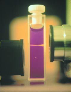
For tissue sections more than ~40 µm thick (intact tissues, whole animals) 2P microscopy is likely to be the only method available to retain sub-cellular resolution. Fluorescent emissions are normally induced by the absorption of a single photon of light shifting the fluorophore to a higher energy state, a photon of a longer wavelength (lower energy) being emitted when the molecule drops back to a lower state. Thus in a confocal microscope a laser, emitting light in the blue/green region, generates fluorescent emissions in the green/red range. An exception to this rule occurs if two photons strike and are absorbed simultaneously, delivering double the energy, resulting in emitted light of shorter wavelength than the excitation light. In a 2P microscope, visible light fluorescence is induced using a near infrared (NIR) laser, yielding two main advantages. Benign NIR photons can penetrate deeper into a specimen. Also since two photon absorption events are very rare, fluorescence only occurs at the exact point of focus where the excitation intensity is highest, reducing background light (See Figure 1.)
Imaging Speed
The necessity for mechanical beam scanning with the confocal technique means that it is at a disadvantage compared to CCD camera systems in terms of image capture rate. Cameras (e.g. the Princetona I-PentaMax, with virtual-chip option, can capture a 160×160 pixel image at 141 frames per second (fps). The Hamamatsub C4880- 81 can similarly achieve 102 fps (164×123). Specialised camera systems such as Redshirt Imaging’sc NeuroCCD-SM can reach 2000 fps (80×80). Using bi-directional scanning, and limiting the scanning area (or resolution) to a 64×256 pixel box, current generation confocals can sustain ~25 fps. Beyond these speeds the point-scanning systems have very small pixel dwell times – low signals, and the detector integration electronics is severely challenged to maintain pixel registration within the imaged plane. However, by reducing the image to a single line, point-scanning laser systems (confocal and 2P) can generate rapid data acquisition from fluorescence indicators, most achieving a scanning rate of 488 lines per second (lps). To attain adequate speed most studies of calcium sparks have been reduced to using this line scan mode.
One way to increase full image capture rate is to scan multiple sets of spots simultaneously over the specimen with a corresponding set of multiple pinholes, an approach which requires the use of a full field detector such as a CCD camera rather than a PMT. Both Perkin-Elmer and VisiTech International supply multi-point scanning confocal systems based upon a rotating microlens array produced by the Yokogawa Electric Corp. In theory, since the array spins at 360 Hz, image capture rates of 360 fps could be achieved although current commercially available systems are limited to around 50 fps by the capabilities of the supplied cameras. It is worth noting, however, that the microlens array and pinhole array are only optimal for 100x objective lenses and that the achievable axial resolution (optical sectioning capability) of the multi-point scanning approach is less than point-scanning confocal microscopes. Stefan Hell has demonstrated a similar rotating microlens system, using a 2P laser source (Straub et al, 2000). The primary advantage here is obtaining better background rejection while not compromising the emission collection to the CCD camera. In addition, the choice of objective lens is not restricted by pinhole matching requirements.
Invasiveness/system sensitivity
CCD camera detection still yields the most photon efficient system for fluorescence microscopy since each CCD pixel collects light during the entire exposure period, permitting lower excitation intensities that can be produced by xenon lamp/monochromators, or even LEDs rather than lasers. Cameras with combined image intensifiers such as the Princetona I-PentaMax or the new Photometricsa Cascade (which incorporates electron amplification within the CCD) have the capability of detecting single molecule fluorescence.
The sensitivity of the confocal microscope is inevitably lower than camera systems since the photon efficiency of the PMT detectors is half (or less) of good CCD cameras. In addition, the emission light must traverse the scanning system optics, folding mirrors, and pass through the confocal pinhole (only 85% of light within first Airy disk). To offset these losses and to generate enough photons to produce a statistically significant number in the short pixel dwell time bright laser sources are required. These high intensities can produce phototoxicity in unstained cells and photo-bleaching in stained cells. This combination tends to limit the viewing time of live cells using UVA/ violet/blue excitation wavelengths.
Although 2P average laser intensities are dramatically higher (~500 times) than confocal laser sources, the NIR photons are lower in energy, and do not seem to perturb transparent cells and tissues with normally used average power levels (<100 mW). The main limitation for live cell imaging seems to be the UVA/violet/blue excitation events, and here 2P imaging produces the smallest excitation volume of all three techniques (see Table 1). Using an excitation wavelength above 1µm – with enhanced detection – has resulted in dramatic improvements in long-term, live cell embryo imaging (Squirrell et al, 1999). The 2P technique also permits the option to bypass the losses associated with the confocal’s scanning optics and pinhole. In addition, these detection- enhanced systems are much less sensitive to focal plane mismatches between excitation and emission wavelengths. Another enhancement possible is collecting photons from the condenser lens, which can double the efficiency to nearer the sensitivity of camera systems.

Don’t forget the lens
The correct choice of microscope objective lens remains an important factor. Generally speaking, the higher the numerical aperture (NA) of a lens the greater its light collection efficiency with significant effects on sensitivity. Short working distance oil or water immersion lenses have a greater NA than air lenses, or long working distance water dipping lenses. Plan Apochromat objectives should be used wherever possible, resorting to Plan Fluor lenses only if UV transmission is required. Water-immersion lenses are preferable for cells/tissues in water beyond 10 µm, water-dipping beyond 200 µm.
And software too …
Similarly, the computer software used to operate the microscope and to analyse the resulting images must be taken into account. As is often the case for modern laboratory instruments, the reliability and/or limitations in this software can often plague otherwise well designed systems. It is unwise to assume that the software package provided by the microscope supplier is capable of the specific forms of image analysis required. Most systems, for instance, have the capability of capturing series of optical sections forming 3 dimensional data sets, but usually have only a rudimentary capability for the analysis of such data. It is worth ascertaining this at the outset and if necessary including third party software with appropriate capabilities (e.g. Bitplane Imaris (*1) or Improvision’s Volocity (*2) for 3D volume display/ analysis, or Universal Imaging’s Meta-Morph (*3) for general purpose 2D time series analysis).
The cost and difficulty of implementing and maintaining the system also needs consideration. It has to be said that many problems remain in the practical application of 2P microscopy in the working laboratory. The pulsed NIR lasers, as well as being costly, continue to be more difficult to set up and maintain in operational order than the visible lasers used in confocal microscopy. The configuration of many early commercial 2P microscopes were often sub-optimal, failing to take full advantage of the new method, particularly in terms of their light detection systems.
In summary, camera systems are simpler, faster, and less expensive, and may be adequate in single cell studies if the fullest sub-cellular detail is not required. The single-point scanning confocal microscope remains ideal for fluorescence imaging of single cells and there are numerous suppliers. Deeper imaging (40-500 µm) requires 2P with its attendant costs and problems. Fast confocal imaging can be achieved using the multi-point scanning systems, at the expense of some spatial resolution and image contrast (a 10% background is always present).
References
Straub M, Lodemann P, Holroyd, Jahn R, Hell SW (2000). Live cell imaging by multifocal multiphoton microscopy. Eur J. Cell Biol. 79, 726-34.
Squirrell JM, Wokosin DL, White JG, Bavister BD (1999). Long-term two-photon fluorescence imaging of mammalian embryos without compromising viability. Nat. Biotechnol. 17, 763-7.
*1 www.bitplane.com
*2 www.improvision.com
*3 www.universal-imaging.com