
Physiology News Magazine

Lab profile: The Biomedical Research Centre, University of East Anglia
Membership
Lab profile: The Biomedical Research Centre, University of East Anglia
Membership
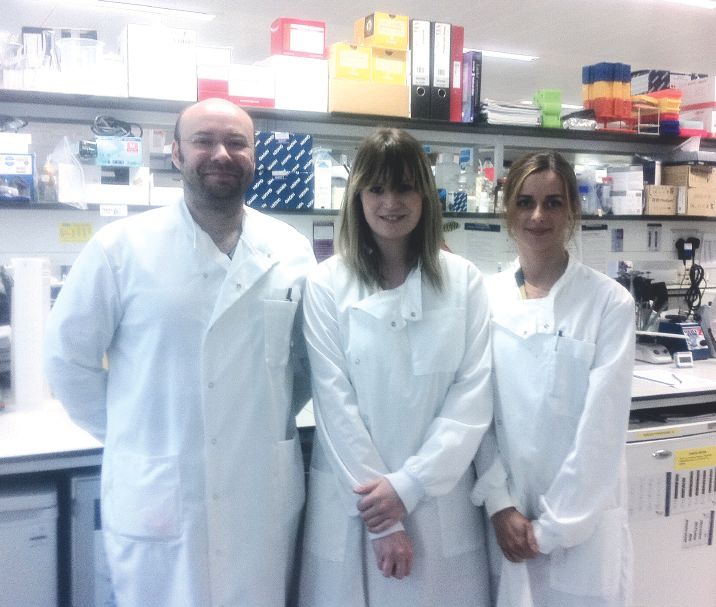
Louise Cully, PhD student, on a lab ‘in its infancy’, benefiting from being located within the Norwich Research Park
https://doi.org/10.36866/pn.88.46
The Univeristy of East Anglia Biomedical Research Centre (BMRC), in which I have been working as a PhD research student since last October, is an integral component of the Norwich Research Park (NRP). The NRP is a collaborative partnership between the University of East Anglia, the Norwich and Norfolk University Hospital, and four independent, world-renowned institutes of research: The John Innes Centre, which focuses primarily on plant genetics and microbiology; The Institute of Food Research; The Genome Analysis Centre; and The Sainsbury Laboratory. Unique multidisciplinary research is made possible through close cooperation between many of the individual research centres located within the NRP’s 160 hectares.
The BMRC is home to over 100 scientists in numerous research groups undertaking pioneering work into arthritis, cancer, cardiovascular and neurological disorders, diabetes and infectious diseases. The BMRC’s Disease Modelling Unit allows transgenic, knock-in and knock-out model systems to be utilised in investigative research, encouraging interdisciplinary coordination from molecular biology up to whole-system physiology.
In 2009, my primary supervisor, Giles Watts, who is currently a lecturer in cell biology and biochemistry within the Faculty of Health, relocated to Norwich following a position as Instructor in Paediatrics and Medicine at the Children’s Hospital in Boston, Massachusetts. His research is conducted around the autosomal-dominantly inherited multisystem disorder Inclusion Body Myopathy associated with Paget’s disease of Bone and Fronto-temporal Dementia (IBMPFD; OMIM to #167320). In 2004, using a candidate gene approach, he identified mutations in valosin-containing protein (VCP) as the causative factor in the pathological development of IBMPFD. It was after this discovery that his area of interest shifted towards the elucidation of the physiological role of normal and genetically mutated VCP in biological tissues; mainly bone, muscle and brain – the three primary tissues affected by VCP mutation.
VCP is a ubiquitously expressed member of the AAA+ (ATPase associated with diverse cellular processes) protein family, with multifarious functions in the ubiquitin–proteasome system (UPS) and retro-translocation of endoplasmic reticulum- associated degradation (ERAD) substrates. VCP binds various combinations of primary and secondary co-factors to form a molecular chaperone complex, shuttling misfolded cytosolic protein aggregates to the proteasome. When a loss-of-function mutant VCP is expressed, cytosolic and nuclear accumulation of aggregated, ubiquitinated proteins is observed. The presence of these ‘inclusion bodies’ and rimmed vacuoles in brain and muscle tissue are some of the diagnostic markers of IBMPFD. The muscle pathology – my own area of research – is accompanied by muscle fibre degeneration, rimmed vacuole formation and ubiquitin-containing sarcoplasmic inclusions. Moreover, muscle fibres of VCPR155H/+ knock-in mice display severely swollen and abnormal mitochondria, suggesting that they may have impaired energy metabolism.
Since joining the Watts lab, my work has been focused on analysing the functional effects of mutant VCP transfection into stable cell lines. To date I have used plasmid expression vectors carrying VCP R155H and A232E mutations in cultured HeLa cells in order to determine the effects of VCP mutant expression on mitochondria dynamics and metabolism. Over the last few years, work on PINK1 and Parkin have highlighted the importance of the UPS in regulating the biogenesis and clearance of dysfunctional mitochondria as part of cellular homeostasis. Since VCP in an integral part of the UPS we believe it is involved in mitochondrial protein ‘quality control’.
My research also involves assessing the physiological and functional effects of VCP mutation in skeletal muscle using a murine VCPR155H/+ model. Under the guidance of my secondary supervisor, Gabriel Mutungi, I am assessing the fatigue characteristics of the extensor digitorum longus and soleus muscles, which consist of mainly type IIb/x fibres and type I and IIa, respectively. This allows us to characterise the fibre type- specific properties of the mutant mice in the context of ageing.
My colleague, Milka Budnik-Zawilska, a second-year PhD research student in the Watts lab, is investigating the role of VCP in the autophagic pathway. As Paget’s disease of bone is one of the pathologies of VCP mutation, Milka’s work involves determining the mechanism by which VCP mutations result in irregular and disorganised bone remodelling, a hallmark feature of PDB due to excessive osteoclast activity. Pagetic osteoclasts are excessively large, multinucleated and contain VCP-, p62- and ubiquitin-positive inclusion bodies, implying a homeostatic role for VCP in the regulation of osteoclast activity in the disease-free state.
While our lab is still in its infancy, we are exceptionally lucky to be part of the BMRC and to work alongside many knowledgeable research scientists with varying areas of expertise, allowing for integration, collaboration and the continuation of high standards of scientific research.