
Physiology News Magazine

Movement automaticity shows less activation, but more connectivity: a model for brain efficiency
Functional MRI (fMRI) was used to investigate interactions in the brain when movements were sufficiently practiced as to become automatic. When a motor task achieved automaticity a group of brain regions became less active, but more strongly connected. Tao Wu and colleagues speculate that this increase in connectivity reflects more efficient brain function when a task is well learned
Features
Tao Wu (1), Piu Chan (1), Mark Hallett (2)
1: Beijing Institute of Geriatrics, Department of Neurology, Key Laboratory on Neurodegenerate Disorder of Ministry of Education, Xuanwu Hospital, Capital Medical University, Beijing, China
2: Human Motor Control Section, Medical Neurology Branch, National Institute of Neurological Disorders and Stroke, National Institutes of Health, Bethesda, MD, USA
https://doi.org/10.36866/pn.73.23
After a great deal of practice people can perform some movements automatically. Automaticity implies that movements can be performed without attention being clearly directed toward the details of the movement. Previous functional neuroimaging studies, including our own, have found that the process of automaticity is accompanied by a reduction of brain activation in several regions, like the cerebellum, premotor area (PMA) and dorso-lateral prefrontal cortex (DLPFC) (Wu et al. 2004; Poldrack et al. 2005). However, the physiology of automaticity was still far from being understood. A further obvious question that arises from these observations is how people can use less brain resources yet perform motor tasks better. We hypothesized that the acquisition of automaticity is not only related to the changes of the magnitude of neural activity, but is also associated with a modification of the interactions within brain networks. In recent years, a great effort has been made in exploring inter-regional connectivity in a given task, which is usually characterized in terms of functional connectivity or effective connectivity (Friston et al. 1993). Effective connectivity implies an interaction between brain regions that might be responsible for a behavioural change. In the current study, we used functional MRI (fMRI) and effective connectivity to investigate the interactions among brain regions when movements become automatic.
Healthy volunteers were asked to practice a sequential finger movement, and after extensive training they performed the task automatically. Automaticity was evaluated by having subjects perform a secondary task simultaneously with the sequential movement. No deterioration in performance indicates that the behaviour is automatic. fMRI data analysis was performed with SPM2 software (Wellcome Institute of Cognitive Neurology, London, UK). First brain activations during the novel and the automatic stage were calculated and compared. fMRI results showed that the pattern of brain activity while performing sequential movement was similar at the novel and the automatic stage, but that the bilateral cerebellum, bilateral PMAs, bilateral parietal cortex, left DLPFC, pre-supplementary motor area (pre-SMA), cingulate motor area (CMA), precuneus, and left putamen were less activated as the sequential movement became automatic. Then, automaticity-dependent changes in effective connectivity were assessed using a psychophysiological interaction (PPI) model (Friston et al. 1997). PPI is defined as the change in contribution of one brain area to another due to a change in experimental condition or psychological context, and aims to explain regionally specific responses in terms of the interaction between the psychological variable and the activity in a specific index area. We chose the left primary motor cortex (M1), bilateral dorsal PMA, bilateral DLPFC, bilateral cerebellum, left putamen, SMA, CMA, and precuneus as index areas because these regions may be involved in the process of automaticity or are important in motor learning. We found that the bilateral cerebellum, CMA, pre-SMA, and left putamen have stronger interactions with a number of brain regions at the automatic stage compared to the novel condition (Fig. 1 and Table 1). In contrast, the precuneus has decreased effective connectivity at the automatic stage.
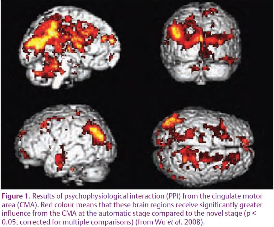
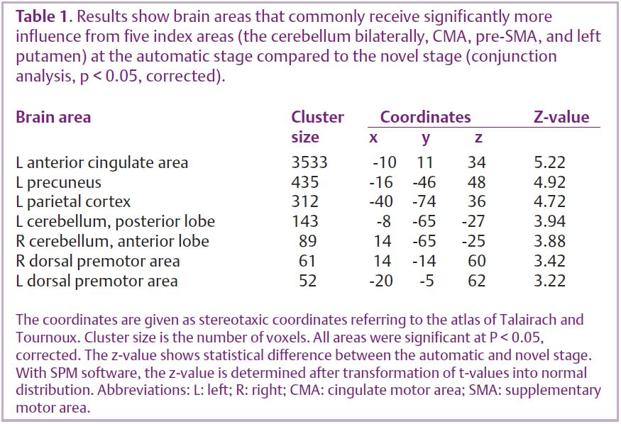
These findings suggest that the process of automaticity is accompanied by a strengthened interaction within much of the central motor networks even though the magnitude of the activation is decreased. With automaticity, brain regions become less active, but some of them increase their effective connectivity. If a movement becomes automatic, then learning must occur, and thus synaptic strengths must change. These changes appear to allow the brain to function more efficiently for the given task, even with a reduced level of activation. The network that does become more connected includes the basal ganglia and cerebellum. In contrast, some cortical regions, like the DLPFC, PMA, and M1, did not show stronger automaticity-related effective connectivity. The primary motor cortex itself is not a part of the automatic networks, perhaps indicating that at this stage it is acting largely in execution mode, carrying out the directions sent to it. The result showing the decreased connectivity of the precuneus can be interpreted that the importance of the cortical attention network decreases when movements become automatic. These findings taken all together provide evidence for the previously poorly supported, but widely held, view that the execution of automatic movements is shifted more subcortically.
The brain is constantly learning new things, but cannot continuously increase its activity. Increasing the functional strength of connections is a solution to this problem and may be an important generalizable property of brain function.
References
Friston KJ, Buechel C, Fink GR, Morris J, Rolls E & Dolan RJ (1997). Psychophysiological and modulatory interactions in neuroimaging. NeuroImage 6, 218–229.
Friston KJ, Frith CD & Frackowiak RS (1993). Time-dependent changes in effective connectivity measured with PET. Hum Brain Mapp 1, 69–80.
Poldrack RA, Sabb FW, Foerde K, Tom SM, Asarnow RF, Bookheimer SY & Knowlton BJ (2005). The neural correlates of motor skill automaticity. J Neurosci 25, 5356–5364.
Wu T, Chan P & Hallett M (2008). Modification of the interactions in the motor network when a movement becomes automatic. J Physiol 586, 4295–4304.
Wu T, Kansaku K & Hallett M (2004). How self-initiated memorized movements become automatic: a fMRI study. J Neurophysiol 91, 1690–1698.