
Physiology News Magazine

Neural WiFi
A new form of communication in the brain by electric fields
Features
Neural WiFi
A new form of communication in the brain by electric fields
Features
Dominique M Durand, Case Western Reserve University, Ohio, USA
https://doi.org/10.36866/pn.116.34
The current thinking about spontaneous brain rhythms is that they result from the interaction between the intrinsic properties of individual neurons and their extrinsic interactions via classical chemical or electrical synaptic transmission. Therefore, extracellular electric fields and voltages recorded in neural tissue are thought to be only a reflection of the underlying neural activity. While non-synaptic effects by electric fields (i.e. ephaptic mechanisms) have been suggested to play a role in modulating ongoing activity, these effects are thought to be reasonably limited, at least during physiologically relevant activity (Anastassiou et al., 2011), but this may not be so.
Neural communication without synapses
While carrying out experiments to study the role of synaptic transmission in the propagation of signals in the brain, we noticed something odd and unexpected. After blocking synaptic transmission by removing extracellular calcium from the environment of the cells, we observed that spontaneous neural events were still propagating. The speed of this propagation was quite fast, around 0.1 m/s. This speed was too fast for diffusion of neurotransmitters and/or ions. Another possible mechanism was the presence of electric synapses (also known as gap junctions), whereby neighbouring cells’ cytoplasms are electrically conductively linked through connexins to form intercellular short circuits that achieve signalling more rapid than that of chemical synapses. However, additional experiments blocking these synapses revealed that those same spontaneous neural events still propagated throughout the tissue (see Fig. 1A) (Chiang et al., 2019; Qiu et al., 2015; Shivacharan et al., 2019; Zhang et al., 2014).
At that point in our research we were left wondering how these events could propagate. After eliminating the usual suspects, we had no choice but to entertain the only remaining viable possibility: electric fields generated during cell activation could recruit neighbouring neurons. This idea was highly controversial since the latest experiments indicated that electric fields could modulate neural activity but were too small to excite neurons by themselves. To convince ourselves that electric field coupling could actually explain our experimental results, we conducted several computer simulations with electric field as the only means of communication between neurons. Computer models did indeed show that electric fields can be sufficient to mediate propagation within the neuronal tissue. However, simulations only show what is theoretically possible and experiments must be designed to directly test the mechanism. We therefore adapted an old method known as voltage clamp to this problem by creating an electrical field clamp system, whereby measurements from detectors sensing the electric field within the tissue were used to generate its corresponding cancelling anti-field that can set and hold (i.e. clamp) the electrical field to zero. This clamp, applied locally was able to block the propagation of the events exactly at that site. Notably, this neural activity was not only eliminated, but could also be modulated or even regenerated by imposed electrical fields. In particular, the application of electric fields with amplitudes in the observed range of the endogenous fields (1 to 5 mV/mm) to the tissue could control the speed of the propagation (Chiang et al., 2019). Therefore both the experiments and simulations confirm that electric field coupling between neurons plays a crucial role in mediating non-synaptic signal propagation.
Self-propagating waves
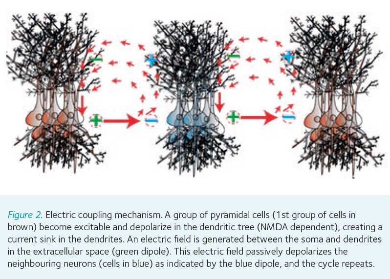
It is well known that action potentials self propagate along axons at speeds determined by the diameter of those axons. However, brain waves recorded during neural activity are not thought to be self-propagating and rather viewed as simply reflections of the underlying neural activity generated by neuronal firing and synaptic transmission. However, our experiments reveal the presence of neural waves that are indeed self-propagating. They maintain their amplitude within the tissue in the absence of synaptic transmission or gap junctions. The speed is also constant at about 0.1 m/s. These data therefore suggest that endogenous electrical fields generated by neural firing in a group of neurons are sufficient to excite their neighbors and generate a wave of activity (see Fig. 2). This hypothesis also implies that endogenous electrical fields can excite other neurons. Stimulation experiments with electrodes placed to generate electrical fields similar to those observed experimentally were indeed observed to induce a propagating wave at the same characteristic speed (Chiang et al., 2019).
Ephaptic effect in sleep waves
This newly found form of communication was discovered while studying the propagation mechanism of brain waves similar to those generated during sleep (Sanchez-Vives and McCormick, 2000). These waves are observed during slow wave sleep and they can be generated in an ex vivo preparation of mouse hippocampal slices, where the waves propagate at the same speed reported above (0.1 m/s). These waves are associated with the regulation of synapses that have been potententiated during wakefulness. In particular these waves are thought to be involved in downregulating synapses that have a low synaptic weight and consolidating the synapses with high weight (i.e. the strength or amplitude of a connection between two neurons). These “sleep” waves were shown to propagate non-synaptically and were not blocked by either presynaptic junction blockers or gap junction blockers (Chiang et al., 2019). Therefore, our results suggest that neural activity during slow wave sleep may regulate synaptic weight by a process involving electric field coupling, a non-synaptic process that would not require presynaptic input into the synapse thereby avoiding generating new potentiation of existing weights.
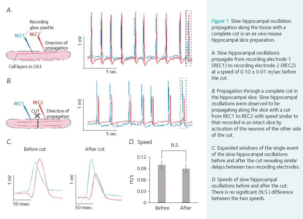
Propagation through a complete cut of the tissue
All along we have been trying to design new experiments to disprove this hypothesis of electric field coupling, without success. Indeed, the idea that electric field coupling, or ephaptic coupling, could allow communication between neurons was met with some resistance and incredulity on the part of other scientists and reviewers. That’s not possible! Did you try this or that experiment? Finally, we predicted that if our hypothesis is correct, then propagation should go through a complete cut of the tissue since coupling by electric fields is carried without any delay through volume conductors. This experiment would cut axons and eliminate gap junctions and synaptic transmission simultaneously. Ex vivo experiments carried out in hippocampus slices confirmed that indeed waves can travel across a physical void in the neural tissue as long as the gap is not greater than 400 µm, at which point the field strength would be too weak due to the inverse-square law (Chiang et al., 2019; Shivacharan et al., 2019) (see Fig. 1 B-D). Similar preliminary results were obtained in mice in vivo and neural activity was observed to cross through a complete transverse cut of the hippocampus. These results indicate that endogenous fields thought to be too small can indeed be large enough to excite other neurons located close by. In addition, the results indicate that the endogenous electric fields can no longer be considered simply as a passive reflection of the underlying neural activity and should instead be thought of as active players in and contributors to neural function.
Endogenous fields can recruit and synchronize neurons in epilepsy
Epilepsy is characterized by increased neuronal firing and hypersynchrony of neural activity. Our recent experiments have implicated these endogenous electrical fields in recruiting neurons during seizures without any involvement of synaptic transmission (Chiang et al., 2018; Chiang et al., 2019). Seizure-like activity was observed to propagate through a cut of the tissue and the propagation could be blocked by cancelling the tissue electric field locally using the newly developed extracellular field clamping circuit. Therefore, our results indicate that this new form of communication plays a significant role in how seizures recruit neurons and could lead to new treatment modalities for seizure control. In particular these results could explain failures of surgical transections of the brain in some patients with epilepsy.
Why is this important?
Until now there were four known ways that neurons could “talk” to each other in the brain: via synaptic transmission, diffusion, axonal transmission and what are known as “gap junctions” between the neurons. Yet when many neurons fire together they generate weak electric fields that can be recorded with the electroencephalogram (EEG) or within neural tissue such as field potentials. These fields were thought to be too small to contribute to neural activity. However, our recent experiments have shown that these electric fields can excite cells which in turn produce electric fields of their own thereby generating a self-propagating wave of activity. Although waves have been observed before, these experiments are showing that these waves can be self-sustained and propagate on their own without synaptic transmission. The function and role of this self-propagation in physiological and pathophysiological neural activity is still a mystery. However, brain waves such as theta waves or sharp-wave ripples are thought to be involved in memory encoding and consolidation. Information is encoded into a neural circuit by a phenomenon called long-term potentiation, which involves simultaneous firing of both pre- and post-synaptic elements. To trim synaptic weights without encoding new information, a mechanism that does not potentiate synapses is required. Waves propagating by ephaptic coupling could regulate synaptic weight during sleep without the need for presynaptic stimulation, since it would not interfere with the synaptic weights already set in the neuronal dendrites.
References
Anastassiou CA et al. (2011). Ephaptic coupling of cortical neurons. Nature Neuroscience 14(2), 217–223.
Chiang CC et al. (2018). Slow moving neural source in the epileptic hippocampus can mimic progression of human seizures. Scientific Reports 8(1), 1564.
Chiang CC et al. (2019). Slow periodic activity in the longitudinal hippocampal slice can self-propagate non-synaptically by a mechanism consistent with ephaptic coupling. Journal of Physiology 597(1), 249–269.
Qiu C et al. (2015). Can neural activity propagate by endogenous electrical field? The Journal of Neuroscience 35(48), 15800–15811.
Sanchez-Vives MV, McCormick DA (2000). Cellular and network mechanisms of rhythmic recurrent activity in neocortex. Nature Neuroscience 3(10), 1027–1034.
Shivacharan RS et al. (2019). Self-propagating, non-synaptic epileptiform activity recruits neurons by endogenous electric fields. Experimental Neurology 317, 119–128.
Zhang M et al. (2014). Propagation of epileptiform activity can be independent of synaptic transmission, gap junctions, or diffusion and is consistent with electrical field transmission. The Journal of Neuroscience, 34(4), 1409–1419.