
Physiology News Magazine
“Sounding out the students” ultrasound in undergraduate anatomy education
Overcoming barriers to ensure a positive ultrasound learning experience
Features
“Sounding out the students” ultrasound in undergraduate anatomy education
Overcoming barriers to ensure a positive ultrasound learning experience
Features
https://doi.org/10.36866/pn.130.27
Dr Keren Bielby-Clarke
University of Bradford, UK
Dr Pip Garner
University of Bradford, UK
The use of portable, handheld ultrasound equipment is increasing in the teaching of clinical skills across a wide range of different undergraduate programmes. This provides flexibility to demonstrate the essential principles of ultrasound in a much wider range of situations than traditional clinical devices. There are clear benefits for undergraduate students, ranging from the hands-on application of anatomical knowledge, development of professional skills in communicating and working directly with patients, and clinical skills in terms of the use and interpretation of the ultrasound scans. However, if unrecognised, cultural, social and personal barriers may negatively impact students’ access to these opportunities.
Introducing ultrasound into anatomy teaching
In the spring/summer of 2022, we received funding for three handheld ultrasound probes, and so began a project to integrate hands-on ultrasound sessions in the Clinical Sciences programme at the University of Bradford (Edwards et al., 2023).
This was a perfect opportunity to enhance our provision of patient-facing clinical skills training, as a simultaneous programme restructure meant that new modules were created with a stronger anatomy theme alongside an increased focus on medical imaging. The programme is designed to provide our graduates with a strong science background alongside the personal and professional skills necessary for a wide range of patient-facing healthcare professions. Therefore, the use of ultrasound was seen as an exciting opportunity to engage students directly with volunteer “patients”, while simultaneously enhancing their direct application of anatomical knowledge.
The University of Bradford is unusual in its diversity, with a population of ~70% BAME students (Advanced HE Race Equality Charter) While this diversity and range of cultures, backgrounds and beliefs is certainly a strength, it can also present challenges when students are expected to interact with each other and with patients to undertake physical examinations or to explore living anatomy and clinical imaging. For example, performance anxiety may occur when carrying out clinical examinations with a group of peers.
Cultural or religious beliefs may restrict the study of the human body in specific circumstances, with physical contact being perceived as discouraged or even forbidden. Personal experiences may also dictate a student’s engagement with (for example) a patient of the opposite sex. It is therefore essential to identify and recognise possible barriers, mitigating them wherever possible, to allow students to maximise these learning opportunities.
Integrating ultrasound
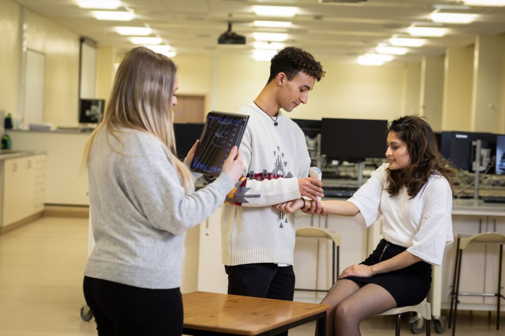
Ultrasound sessions were directly integrated into taught practical sessions to provide opportunities for direct application of anatomical knowledge, in topics such as thoracic, abdominal or pelvic anatomy, the upper and lower limbs, and the back. In planning the sessions, careful consideration was given to the comfort of the patient and students, while maximising group throughput (due to limited practical time available in the timetable, coupled with large cohorts of students). Students were provided with instructions in a worksheet and supported by an academic or a trained demonstrator while carrying out the ultrasound.
From the outset our students demonstrated a wide range of confidence levels and engagement, with some eager to interact with the patient and use the ultrasound, and others remaining distant and silently observing. There are many reasons why students may not engage. We started the sessions early in the academic year, so the students were acclimatising to the course and to each other, and for many of them this would also have been their first experience interacting directly with a patient. For others there may have been other underlying concerns. This evaluation was designed to identify and where possible attenuate these barriers to future experiences.
How do we know what we don’t know?
Informal focus groups were set up to gather opinions and feedback from students across the different cohorts. These groups comprised a maximum of five students per group with one member of staff. They consisted of a directed but informal discussion around specific issues and challenges, as well as positive experiences, when using the ultrasound equipment to work with patients. Since the ultrasound was used in different ways and to different extents across the years of the programme, volunteers were sought from the foundation, first and second year separately.
Within the groups, the main focus of the discussion was to identify any specific benefits that the students noted, or barriers to the use of the ultrasound equipment when interacting with patients in this quasi-clinical setting. The questions to guide the discussion included:
- What were your feelings before, during and after the ultrasound session?
- Were there any barriers to you taking part or conducting an ultrasound examination?
- What could we have done differently to prepare you?
- Was the experience worthwhile?
- How will you approach similar sessions in the future?
These questions were chosen to encourage the students to reflect on their experiences, highlighting what they gained from it as well as any specific issues, and identifying future solutions that may ease the process.
Ultrasound “is a privilege” – but group size is a drawback
Within the focus groups, students were very positive about the overall experience. They highlighted the benefits of applying their taught anatomy knowledge to the real-life patient, exploring the different structures and seeing the actual positions and locations.
“I think an ultrasound is a privilege because you could have just given us images of X-rays”
Large group size is a well-recognised barrier to effective engagement in practical situations such as these, requiring a level of confidence that cannot be assumed, particularly for students in the early stages of their degree. Group size was highlighted by a number of students as a major barrier to engagement, as many did not feel comfortable “performing” in front of their peers. Careful consideration in the future and practice in a low-stakes environment (without a patient) may remove this obstacle. However, working in larger groups allows more reticent students with anxiety around patient interactions to act initially as a bystander to develop their levels of comfort, while the more confident students take the lead.
Surprisingly, when discussing and consolidating the experience there is an almost competitive aspect in terms of locating and identifying the anatomical structures.
“We discussed our findings. Oh, I found this. Did you find that? I saw the striations. Did you? I found a bit of the liver.”
In this way the larger groups enable interaction and engagement even after the session.
It became very clear that there was not enough preparation beforehand in terms of actually using the probe, or interpreting the images in terms of specific anatomical structures and landmarks. However, the presence of the academic demonstrator to guide and question students during the activity was useful to direct attention and check understanding.
Prior notice: preparation is vital
Some of the students reported being “surprised” that they would be working with a patient. However, they then viewed this as them to interact with individuals in a quasiprofessional setting. We also had students who reported feeling under-prepared in terms of how to address and speak to a patient, and how to actually use the ultrasound; for example, there were concerns about causing discomfort by pressing too hard with the ultrasound probe.
“I tried to talk, but I didn’t know what to say and it was a bit awkward. So I didn’t.”
When students are given prior notice of the patient and anatomical areas to be studied, they have time to prepare mentally and also to rehearse professional skills such as basic communication. Simply “dropping” students into a session with no prior warning may leave them uncomfortable and with difficulties engaging. Students would therefore benefit if provided with specific guidance on patient communication, in order for them to become comfortable with their role as a healthcare professional beforehand.
Likewise, examination of a patient’s abdomen or pelvis may require more consideration beforehand, compared to the arm or hand, due to generalised cultural significance of the viewing or uncovering of body regions normally not visible.
Recognising and clarifying religious and cultural barriers
In terms of the cultural or social barriers, there were several different issues highlighted. Alongside the expected gender-based tentativeness (particularly when examining areas of the body that would normally be covered, for example the abdomen or pelvis) there were areas of concern around physically touching another individual’s skin, or of discomfort when exposing parts of the body to colleagues when working in student groups. It was mentioned several times that students were initially concerned about working with a male or female patient particularly, but then realised that in practice, they would be dealing with all patients and so this was a valuable learning experience.
“We saw that there was the patient and it was like, Oh, okay, there’s a patient there. And then the more you thought about it, the more you realise, hang on, that’s what we’re going to have to do in the healthcare setting anyway.”
Significant and specific religious barriers were also identified. In culturally diverse cohorts this can often be an unseen or unrecognised issue, particularly when there is a “mismatch” in student and lecturer demographic, in terms of religion, race or background. Students reported taking it upon themselves to consult religious scholars, in order to confirm that they were permitted to undertake clinical interactions of this nature, given the educational/professional setting and training.
In future, consultation with appropriate religious scholars and faith advisors will be undertaken by academics, ensuring that any student concerns can be answered quickly and effectively to reassure students and remove any perception of impropriety.
“Failing to prepare is preparing to fail”
Thorough preparation cannot be too highly valued, as this avoids time wasted in the ultrasound session and allows students to maximise the engagement with their patient. To this end, carefully structured resources to allow students to check their anatomical knowledge of the region to be examined and explore example ultrasound images to prepare for the practical session will be essential. It is also important to consider the effective timing of the release of these resources, long enough before the session that students have time to engage and explore them, but not so far in advance that they are forgotten by the session itself.
“I think maybe if you had videos showing how to do each ultrasound prior to your session, people would be more comfortable with doing it”
“Having that prior knowledge would help in this session because I’ll know what to look for and be like, Oh, this is how I do it”.
So what’s next?
The main focus of this evaluation was to ensure that students felt prepared and comfortable prior to their interactions with patients, allaying concerns both from a personal and professional perspective, and in terms of their knowledge.
We have identified several points for improvement and clarification, both for provision of information and preparation, but also for guiding and directing our students to specific points of consideration that they may not have otherwise appreciated.
Moving forward, these improvements and additions will enable us to create a safe and engaging space where students can interact and learn without fear of judgement.
References
Edwards H et al. (2023). Assessing student perception of portable ultrasound imaging in undergraduate anatomy education. Clinical Anatomy 36, 742-753.
Advanced HE Race Equality Charter: University of Bradford (Bronze Award)