
Physiology News Magazine

The British Heart Foundation’s Reflections of Research competition
News and Views
The British Heart Foundation’s Reflections of Research competition
News and Views
https://doi.org/10.36866/pn.95.14
The British Heart Foundation (BHF)’s Reflections of Research competition seeks out the most extraordinary and surprising views of the heart and blood vessels amongst the research the charity funds. The annual competition is open to researchers supported by the BHF, and accepts images from all areas of cardiovascular research.
Simon Gillespie, Chief Executive at the BHF, said of last year’s competition:
‘This isn’t just visually arresting art; these pictures are reflections of our life-saving research, which makes them even more beautiful.’
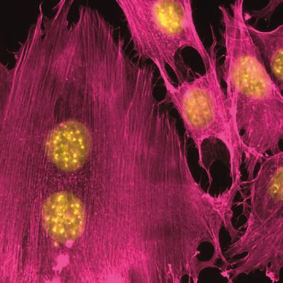
Pink in art
Fluorescence staining of the nuclei (yellow) and actin filaments of endothelial cells (in pink) which provide a defensive wall against toxins to keep blood vessels healthy.
Kenneth Cheung, Queen Mary, University of London, UK
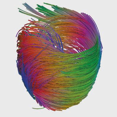
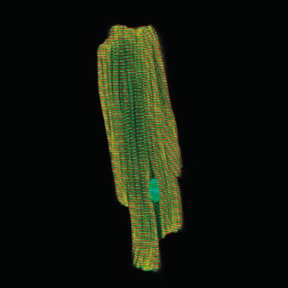
Heart strings
Diffusion Tensor Imaging from an MRI scan of a heart. The tracks show movement of water molecules in the heart muscle, which reveals how the muscle cells are aligned. Resultantly, the streamlines shown in the image represent the orientation of ‘muscle fibres’ in the heart’s left ventricle.
Patrick Hales, University of Oxford, UK
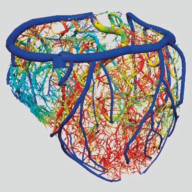
Feeding the heart
This virtual model shows the blood flow through vessels serving the heart.
Nic Smith, Kings College London and University of Oxford, UK
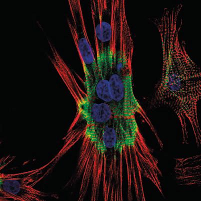
The ‘B’ of the Bang
Cultured cardiomyocytes stained for subcellular structures; nuclei (blue) are surrounded by the cytoskeletal (red) and beating elements of cells (green).
Elizabeth Ehler, King’s College London, UK
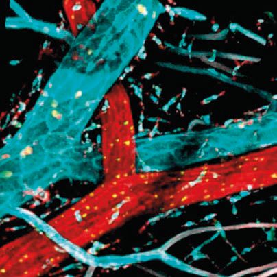
Opposites attract
Blood vessels stained for arterial (red) and venous (blue) markers.
Krishma Halai, Queen Mary, University of London, UK
Pulling together
Confocal image of a mouse cardiomyocyte stained for the nucleus (cyan) and two different proteins, alpha-actinin (red) and titin (green) involved in the contractile function of the cells.
Thomas Cahill, University of Oxford, UK
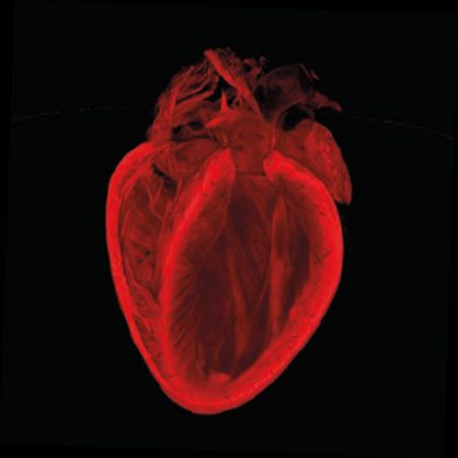
The Broken Heart
3D structure of an adult mouse heart. The imaging technique used here is being developed to allow us to better measure the extent of injury after heart attack, and to assess repair.
Gillian Gray, Megan Swim, and Harris Morrison, University of Edinburgh, UK
The winning images can be viewed online at bhf.org.uk/reflections. You can support the BHF’s life-saving science by donating at bhf.org.uk