
Physiology News Magazine

The horse – the athlete with the ultimate locomotor system
Features
The horse – the athlete with the ultimate locomotor system
Features
Amy Barstow & Renate Weller
Royal Veterinary College, Hatfield, UK
https://doi.org/10.36866/pn.102.26
Horses are exceptional athletes but their anatomical and physiological adaptations which give them a competitive edge simultaneously put them at risk of injury.
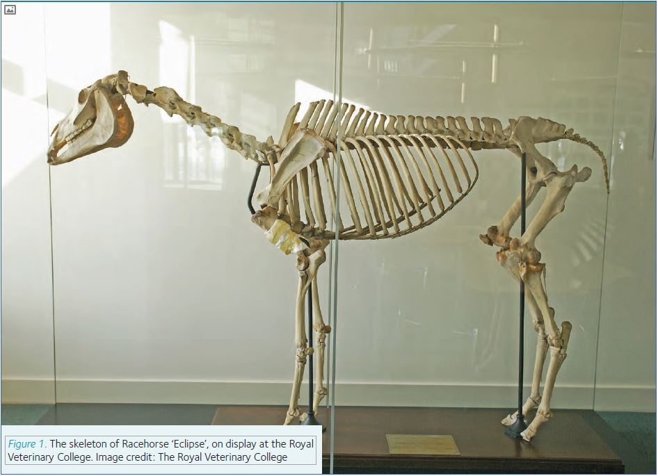
For the majority of us the word ‘athlete’ conjures mental images of the Olympics; Paula Radcliffe, Usain Bolt, Mo Farrah, Dame Kelly Holmes – human athletes. Nowadays, equestrian events might even spring to mind too, following the success of Team GB Riding at London 2012. However, horses in sport are nothing new. The very first official Olympic Games in 778 BC had four equestrian events. Furthermore, the very beginnings of veterinary education in the UK – the founding of the Royal Veterinary College in 1791- is linked to the death of the unbeatable racehorse Eclipse (Fig. 1). His owner asked for a post-mortem examination to find out what made him so fast, resulting in the first post mortem assessment for performance.
A horse’s specialised anatomy makes it suitable for a range of sports: gymnastic dressage divas, high jump show jumpers, agile polo ponies and of course the Ferraris of the horse world, the racehorses. However, you also find the more versatile all-rounders: triathlete eventers competing in dressage, show jumping and cross-country.
Horses and humans are more similar than you’d think!
At first glance, you might think that equine athletes are far removed from their human counterparts and that it would be impossible to draw any comparisons. But you would be wrong. Prior to changing hands (and good horses go for vast sums of money), most horses undergo pre-purchase exams including thorough medical checks, radiographs and – more and more commonly – magnetic resonance imaging (MRI), much like football players. Top human athletes have a support team and so do horses: veterinarians, farriers, physiotherapists, trainers, saddle fitters, nutritionists, grooms and biomechanists work together to ensure optimal performance and to safeguard equine welfare. Like in human sports medicine, veterinary sports medicine strives to be evidence-based and the body of scientific studies on exercise physiology, sports orthopaedics and equipment interventions has been increasing rapidly.
All athletes walk a fine line between being the best and risking an injury and equine athletes are no different. Like injuries to rugby players can reflect the position they play, equine injuries show associations with specific sports (Murray et al., 2006). However, if things do go wrong, unlike humans, horses cannot tell us where it hurts. Ultimately, the same diagnostic techniques are used to investigate poor performance and injuries; however, the fact that horses cannot talk, are somewhat bigger than humans, and have a different anatomical and physiological make-up, means specific adaptations are required in diagnosis and treatment.
As with most things in life, prevention is better than cure and there is extensive research into reducing injury risk in our equine athletes. This not only improves welfare but also optimises their performance as the athletes they truly are.
Hybrid vehicles, pogo sticks and catapults
Horses owe their ability to perform in a number of different sporting activities to their highly specialised anatomy. Their cursorial nature means they are designed to cover vast distances at a slow pace, while grazing for nourishment: a prerequisite for survival on the sparse vegetation of the prairie type land the horse originated from. However, at the same time they have evolved as prey animals with the capacity to make a speedy escape if needed. The horse supports its locomotor requirements by having developed anatomical and physiological features that reduce the metabolic cost of locomotion. The horse has long and light limbs, which is achieved by reducing the number of lower limb bones to one weight-bearing column, and elongating the limb by essentially walking on their tiptoes.
Unlike human legs, that are pretty much straight from the tarsus (ankle) up, horse legs are angled with the metacarpophalangeal joint being the pivot point. This joint is in hyperextension during normal standing and goes into extreme hyperextension during movement (Fig. 2). While it may not seem very advantageous at first glance to have what is essentially an unstable joint, this configuration allows the horse to use its bodyweight to save energy during movement. Long and strong flexor tendons, with properties that make structural engineers jealous, run down the back of horse’s legs and around the metacarpophalangeal joint. During movement, when the body weight compresses the leg, these tendons get stretched and store elastic energy. This energy is returned when the leg leaves the ground, tendons are known to be able to return up to 93% of energy. This mechanism pretty much resembles a pogo stick. At rest, tendons and ligaments contribute to the passive stay mechanism, which allows the horse to stand and sleep with minimal muscular effort.
The horse takes energy saving through tendinous elastic recoil even further. The biceps muscle has a specialised, internal tendon which stores elastic energy slowly, whilst the hoof is in contact with the ground, before quickly releasing its elastic potential energy to rapidly accelerate the forelimb forwards into the swing phase (Wilson et al., 2003). This process quite literally catapults the horse away from danger – or out of the starting gates.
Like hybrid cars, the horse uses metabolic energy (fuel) to feed its muscles to initiate locomotion. The movement of the horse and vehicle itself then drives energy storage mechanisms: elastic potential energy in the horse and the charging of batteries in a hybrid vehicle.
A gambling problem
At the races it’s not just the punters who might have a gambling problem. Horses training at the top of their sport are ‘gambling’ with their anatomy. All the adaptations that make them faster and more efficient also make them more vulnerable to injury. If you want to move your limb fast it needs to be light and you need gracile bones and tendons. Reducing the cross-sectional area of structures makes them more vulnerable to breakage.

We describe the horse as operating at a low safety factor. A safety factor is the load that can be applied to something before it breaks. Things like lifts and stairs operate at a safety factor of at least 10 times the maximal load they are expected to take. Horses, however, operate at full gallop to a safety factor as low as 1.5 times. This is reflected by the fact that approximately 50% of racehorses in training, at any one time, can be affected by injury to their superficial digital flexor tendon (SDFT).
A pain in the leg
If you’ve ever been running and found that your shins hurt afterwards, then perhaps you can empathise with some horses! ‘Bucked shins’, as it is called in horses, is a very common and minor condition that affects racehorses, especially beginners. It is often the result of an inappropriate training regime leading to a shift from adaptive bone remodelling for repair to pathological bone remodelling on the front of the third metacarpal bones. Horses will have painful, hot and swollen metacarpal bones and you can easily feel this with your fingers and quickly make a diagnosis. Rest and modification of the training program is often all that is needed to resolve the problem.
Bucked shins is an example of a minor injury sustained due to repetitive exercise induced damage or cyclical overloading of the musculoskeletal system. Like humans, cyclical overloading of the musculoskeletal system is by far the most common cause of orthopaedic injuries in horses: the rate of exercise induced damage out strips the body’s capacity for repair.
Horses place their limbs under extreme loads. At full gallop, each limb is loaded with approximately 2.5x its bodyweight, that’s 1250kg in an average sized 500kg racehorse. Considering the small cross-sectional area of a horse’s limb, that is an enormous amount of pressure! What’s more is that bones and tendons in horses have been estimated to have a ‘working life’ of as little as 10,000 cycles, before they wear out. A galloping horse will impose about 220 loading cycles per mile and it does not take advanced math skills to figure out that racehorses indeed ‘skirt very close to the edge’. It is therefore no surprise that horses can suffer numerous injuries, which range from the common superficial digital flexor tendon injuries to the less common, more catastrophic injuries, such as stress fractures.
Just tell me where it hurts
As with any athlete, injury is an inevitable part of the game and we owe it to these horses to provide prompt and thorough veterinary attention. Sometimes an injury will be obvious – a wound for example. Other times, it is not, and veterinarians do not have the luxury of being able to ask their patients where it hurts. They must be ‘pain detectives’. Careful palpation of the horse’s musculature, tendons and joints, as well as critical observation of the movement of the horse at walk and trot in straight lines and in circles on both soft and hard surfaces is the usual starting point.
Some problems can only be observed under saddle when a specific movement or task is required of the horse, so observation of the horse during ridden exercise is therefore necessary too.
The first task is to decide which leg is the biggest problem. Since most orthopaedic problems are ‘wear and tear’ issues we often have to deal with multiple leg and multiple site problems. There is a limitation to human’s ability to detect lameness in horses, so sensor-based movement symmetry analysis, as-well as force-plate and pressure mat-based systems can be used in referral centres to aid the veterinarian in this task.
Once we know which leg to start with, diagnostic analgesia is used to localise the area of pain. This involves the ‘blocking’ of specific nerves or synovial structures with injections of local anaesthetic solution. The first block is placed most distally on the limb (typically just above the heel bulbs) and the horse is observed exercising again. If the gait is not improved, another block is placed higher up the leg and so on until improvement is seen. Once an area is identified as the region where the pain is coming from, specific synovial structures within this area – joints, bursae or tendon sheaths – can then be ‘blocked’ with anaesthetic.
Sounds simple right? Not quite; the anaesthetic agents can unhelpfully diffuse to other locations, and in some horses, nerve anatomy varies from the norm, which can be misleading.
Photoshoot
Diagnostic imaging is the mainstay of confirming orthopaedic disease in horses. The methods of imaging are the same as for humans: radiography, ultrasonography, magnetic resonance imaging (MRI), computed tomography (CT) and nuclear scintigraphy.
Radiography and ultrasound are the most commonly used imaging techniques in the veterinary world. They are relatively cheap and can be used in the standing horse in their own home. It is the norm for equine vets to make house calls and horses only get transported to see a veterinarian in more complex referral cases. Ultrasonography is, for example, commonly used to diagnose tendon and ligament injuries, which are especially common in the superficial digital flexor tendon. Osteoarthritis is the most common orthopaedic problem identified on radiographs, however the clinical significance of these findings is often questionable. This often makes interpretation of radiographs performed as part of a pre-purchase exam especially challenging.
Radiography and ultrasonography have their limitations. Some regions of the horse refuse to yield their pathologies willingly. The hoof for example contains many vital soft tissue structures that cannot be visualised radiographically and are not easily accessible with ultrasound as they are enclosed within the dense horn of the hoof capsule. In the forelimb, lameness can be localised within the foot in 80% of riding horses and MRI of this region is hence very common. In the past, horses tended to be anaesthetised and scanned using a human MRI scanner, however, nowadays it is more common today to use a system specifically developed for use in the standing horse. Horses carry a 1% risk of dying under general anaesthesia so veterinarians prefer to perform any procedure in conscious standing horses. Horses are flight animals and many standing procedures require careful sedation on top of expert handling.
Size matters
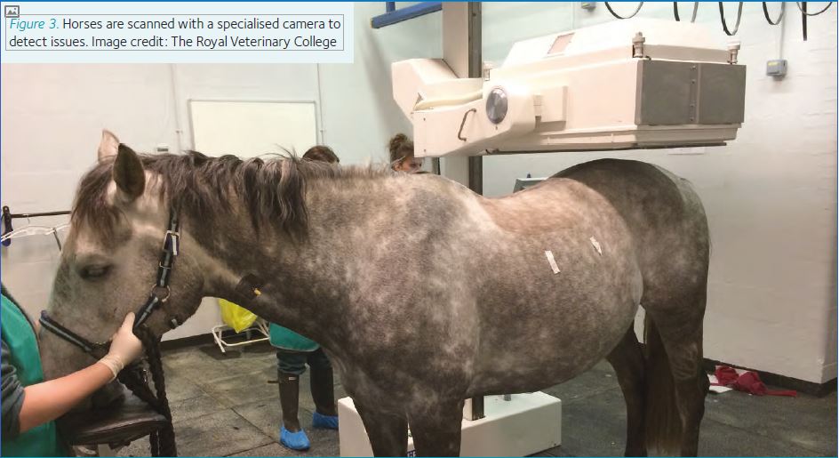
While humans (and dogs) with back problems can benefit from MRI and/or CT evaluation, a normal sized adult horse cannot, they are simply too big to fit in an MRI or CT scanner. Areas like the back and pelvis can be radiographed and ultra-sounded, but the pure size of the horse often makes it difficult to get good quality images, and a technique called nuclear scintigraphy is often employed when bony lesions are suspected. Horses are given an intravenous injection of a radioactive nucleotide (e.g. technetium99) which seeks out bone tissue and accumulates in areas of increased bone metabolism such as a fracture site. The horse is then scanned with a specialised camera (Fig. 3) that detects the radioactive signal and creates an image of nucleotide distribution through the skeletal system. One such example of nuclear scintigraphy use is to diagnose pelvic stress fractures, which occur in horses racing over jumps. Many equine athletes that are presented for a performance issue suffer from multiple problems. It is therefore necessary to employ multiple imaging techniques, based on a thorough clinical investigation, to diagnose the problem(s) the horse has and to treat them accordingly.
Straight from the horse’s mouth – lessons from horses
The high numbers of racehorses suffering from injuries to their SDFT resulted in an intensive search for innovative treatment methods. Tendons heal, but their functional elastic properties are not fully restored as a result of scar tissue formation.
Researchers at the Royal Veterinary College addressed this problem by looking into the application of stem cell therapy in tendon lesions. Mesenchymal stem cells (MSC) are extracted from the sternum bone marrow, and are grown over three weeks before being injected into the SDFT lesion under ultrasound guidance. This technique has been successful in reducing the re-injury rate in horses with SDFT lesions (Godwin et al., 2012).The use of stem cell therapy even features in the rags to riches film ‘Dark Horse: The incredible true story of Dream Alliance’. From this research, the use of stem cell therapy for the treatment of orthopaedic problems has advanced and translated into human medicine, e.g. in the treatment of rotator cuff and Achilles tendinopathies in human athletes.
Silver and Gold
It is of great importance to investigate superior therapeutic techniques for injury management in equine athletes. However, this research is just the tip of the iceberg. Investigation of bespoke surfaces to ride upon, shoes, saddles and bridles for horses to wear, training, feeding and management techniques are just a few examples of current research areas. All focused on improving horse welfare, reducing injury and perfecting performance.
In line with human athletes there is a constant strive to improve equine performance to make the often small difference between winning Olympic silver and winning gold!
Technology Age
The big limitation of working with these speechless animals is that we have limited ways to detect sub-clinical problems. Experienced riders and trainers may detect subtleties in a horse’s performance, such as struggling to turn in one direction compared to the other, not moving as well, having poles down when jumping or not being as fast as they once were. However, small underlying concerns are often not addressed until they are causing much bigger problems in performance. The ability to intervene when an injury is very new, before it causes a significant loss of performance and pain, is essential in optimising welfare and performance. In the technology age, we are working towards improving early diagnoses in a variety of ways.
Biomechanical analysis and exercise physiology assessment of human athletes is common in detecting both underlying problems and driving training programmes. Performance in the horse has been assessed in gait labs for decades. However, recent advancements in sensor-based technologies allows assessment of the horse during sporting activities, and has led to a much more common use of objective parameters (Pfau et al., 2014).
Racehorse trainers, for example, now routinely equip their horses with devices that record heart rate, stride length and frequency, and movement symmetry. Changes in these parameters alert trainers to potential problems and aid in the adjustment of training and racing strategies, shoeing and medication protocols. However, these techniques aren’t restricted to use in the horse, they can be applied to the rider to achieve a team perspective too.
Conclusion
All in all horses are incredible athletes. They would be able to win medals in several human Olympic disciplines should we allow them to compete against humans or other animals and to top it off they would do it with a rider on top! This is in part due to their anatomy but also due to their cooperative and trainable nature. They deserve to have their welfare regarded as of paramount importance, which is only achievable through knowledgeable, multidisciplinary support teams and by furthering scientific research.
References
Godwin EE, Young NJ, Dudhia J, Beamish IC, Smith RKW (2012). Implantation of bone marrow-derived mesenchymal stem cells demonstrates improved outcome in horses with overstrain injury of the superficial digital flexor tendon.
E Vet J 44(1), 25–32
Murray RC, Dyson SJ, Tranquille C, Adams V (2006). Association of type of sport and performance level with anatomical site of orthopaedic injury diagnosis. E Vet J Suppl 36, 411–416
Pfau, T, Spicer-Jenkins C, Smith RKW, Bolt DM, Fiske-Jackson A, Witte TH (2014) Identifying optimal parameters for quantification of changes in pelvic movement symmetry as a response to diagnostic analgesia in the hindlimbs of horses.
E Vet J 46(6), 759–63