
Physiology News Magazine

Tremor
Bettina Pollok and colleagues consider what is physiological in pathological tremor. As with much else, networks are the key
Features
Tremor
Bettina Pollok and colleagues consider what is physiological in pathological tremor. As with much else, networks are the key
Features
Bettina Pollok, Joachim Gross & Alfons Schnitzler
Department of Neurology, Heinrich Heine University, Duesseldorf, Germany
https://doi.org/10.36866/pn.57.26
Since the pioneering neuroanatomical studies of Paul Broca (1824-1880) and Carl Wernicke (1833-1904), it is generally accepted that different brain areas are associated with specific functions. However, functional neuroimaging techniques demonstrate that even the execution of apparently simple tasks is associated with neural activity in a widely distributed cerebral network comprising cortical as well as subcortical structures. Although these studies associate certain tasks with activation of specific brain structures, it remains unclear how information is interchanged between these areas.
There is growing evidence that synchronization of oscillatory neural activity might be a fundamental mechanism of information coding in the brain (Singer, 1999). Coherence and phase synchronization are established measures for quantifying coupling between different brain sites. Recent studies substantiate the hypothesis that relevant information in the brain is coded by accurate timing of neuronal discharges in large scale networks. Such synchrony between spatially distributed neural activity can be investigated non-invasively by using magnetoencephalography (MEG) together with a recently developed analysis tool Dynamic Imaging of Coherent Sources (DICS; Gross et al. 2001). MEG allows for the investigation of brain activity in the range of milliseconds, which is a fundamental premise for characterizing synchronized oscillatory activity. Additionally, DICS provides a topographic map of coherence between different brain sites associated with the execution of a specific task or with a specific motor behaviour. Thus, MEG data provide information about brain structures involved in a task and –most importantly – about functional interactions between these areas. Since it has been argued that relevant information in the brain may be coded by changes in the dynamic interplay between different structures without changes of the local activity, investigation of the dynamic interaction between neural assemblies reveals new insights into the understanding of how information is processed in the brain.
In our recent work we have focused on the investigation of synchronized oscillatory activity associated with pathological as well as physiological motor behaviour. Tremor is defined as an involuntary oscillating movement of a body part, most frequently the upper extremities. It can be observed as a physiological phenomenon in healthy volunteers and as a pathological symptom associated with a wide variety of movement disorders. As early as 1886 Horsley and Schafer speculated that tremor might have – at least in part – a neurogenic basis (Horsley & Schafer, 1886). In a recent study it has been demonstrated that resting tremor in Parkinson’s disease (PD) is associated with a cerebello-diencephalic-cortical network oscillating at tremor as well as at double tremor frequency (Timmermann et al. 2003). These data agree well with the assumption that central mechanisms play a crucial role in the origin of tremor. However, the results raise the question about the specific pathological nature of the demonstrated network: does it reflect a pathological phenomenon per se, or does it represent a functional network underlying physiological motor behaviour, which may be specifically altered in PD resulting in resting tremor?
To answer this question we investigated cerebro-cerebral coupling in a group of healthy subjects voluntarily imitating the typical antagonistic resting tremor (Pollok et al. 2004). Our results demonstrate that, indeed, the same oscillatory network subserves voluntary tremor as well as involuntary resting tremor. Our data, therefore, indicate that coupling within cerebral networks represents a fundamental characteristic of motor control, and that pathological movements like PD tremor may be due to alterations within such physiologically pre-existing networks. Figure 1 summarizes the localization of coherent activity in one representative subject imitating the typical antagonistic PD tremor.
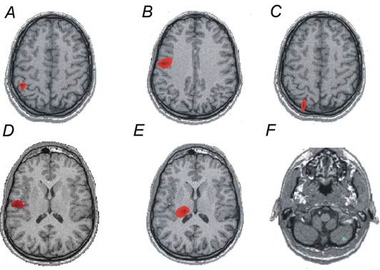
The constituents of the oscillatory network of pathological PD and voluntary tremor are the same.
However, we found important differences when comparing coupling strength within this network between PD patients and healthy subjects imitating resting tremor. First, significant coupling between the diencephalic structure and the primary sensorimotor cortex was reduced in the healthy controls as compared to the PD patient group. This implies a stronger influence of the thalamus on the activity of the primary motor cortex in PD patients. Second, coupling between the premotor cortex and the primary sensorimotor cortex was enhanced in the healthy group most likely indicating that the premotor cortex drives M1 resulting in the voluntary 3-6 Hz tremor. In contrast, in the patient group M1 might be driven by deep diencephalic structures like the thalamus, resulting in involuntary tremor.
Although one has to be cautious when comparing data from patients with those from healthy subjects, since subjects and patients were not age-matched, our data substantiate the hypothesis that PD tremor is based on a physiological oscillatory network, and that characteristic alterations within this network are most likely associated with the generation of tremor.
To summarize, the investigation of oscillatory interactions between brain sites involved in a specific task provides physiologically and pathologically important insights into the functional connectivity between brain areas. The comparison between healthy subjects imitating tremor and PD patients clearly demonstrates that it is not necessarily the local activity of brain areas, but rather the dynamic interplay between these structures, which might be crucial for the understanding of physiological and pathological motor behaviour.
References
Gross J, Hämäläinen M, Timmermann L, Schnitzler A & Salmelin R (2001). Dynamic imaging of coherent sources: Studying neural interactions in the human brain. Proc Natl Acad Sci 98, 694-699.
Horsley V & Schafer E (1886). Experiments on the character of the muscular contractions which are evoked by the excitation of the various parts of the motor tract. J Physiol 7, 96-110.
Pollok B, Gross J, Dirks M, Timmermann L & Schnitzler A (2004). The cerebral oscillatory network of voluntary tremor. J Physiol 554, 871-878.
Singer W (1999). Neuronal synchrony: a versatile code for the definition of relations. Neuron 24, 49-65.
Timmermann L, Gross J, Dirks M, Volkmann J, Freund HJ & Schnitzler A (2003). The cerebral oscillatory network of parkinsonian resting tremor. Brain 126, 199-212.