Background:
Electrophysiological techniques have been widely used to study the functional mechanisms of the brain’s sensory-motor networks in both health and neurodegeneration. However, such assessments of spinal cord activity remain highly challenging and underexplored. This gap is concerning, considering that cervical spinal degeneration[1] is a key clinical indicator of disease severity and future disability in neurological conditions such as Multiple Sclerosis (MS)[2].
To address this gap, our team recently developed a standardised high-density electrospinography (HD-ESG) system (SC10X/U)[3], an up to 76-channel platform designed to reliably record spinal cord electrical activity and enable assessment of spinal function. This is especially relevant in conditions like MS, which features a clinico-radiological paradox, i.e., changes in structural spinal MRI measures, such as atrophy, fail to predict or correlate with disability progression despite strong clinical evidence of spinal involvement.
Objective:
Here, we used HD-ESG to quantify signatures of spinal High-frequency oscillations (HFOs) in response to the median nerve stimulation (MNS), in MS. HFOs are a measure of localised synchronous presynaptic neural activity[4], [5], reflective of functional integrity. To our knowledge, this is the first study to investigate spinal-HFO responses in people with MS (pwMS).
Methodology:
64-channel ESG[3] was recorded in response to the stimulation (right wrist, 1.5 X Motor Threshold, 2Hz) from 14 pwMS and 18 controls. A total of 1400 evoked responses were recorded per participant. Signals were pre-processed and bandpass filtered between 350- 1400 Hz. The evoked HFO (phase-locked) responses were obtained by averaging the resulting signals across all responses.
Temporal HFO characteristics were compared between groups using the Mann-Whitney U test with adaptive false discovery rate (q = 0.05). Characteristics include: onset latency, peak latency, and normalised peak amplitude (Peakamp).
Results:
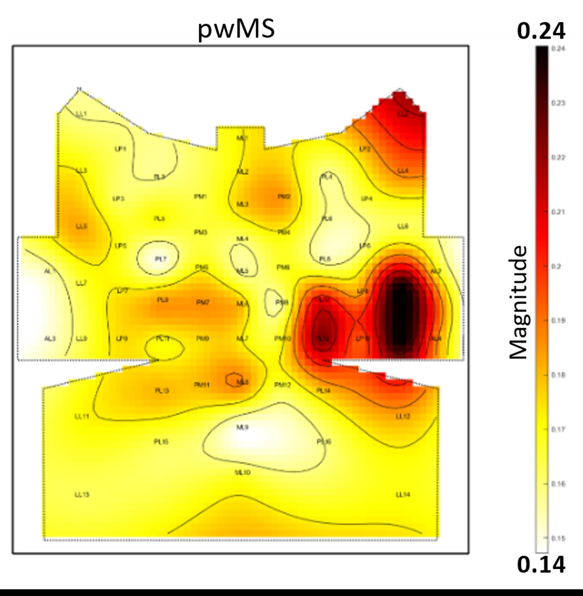
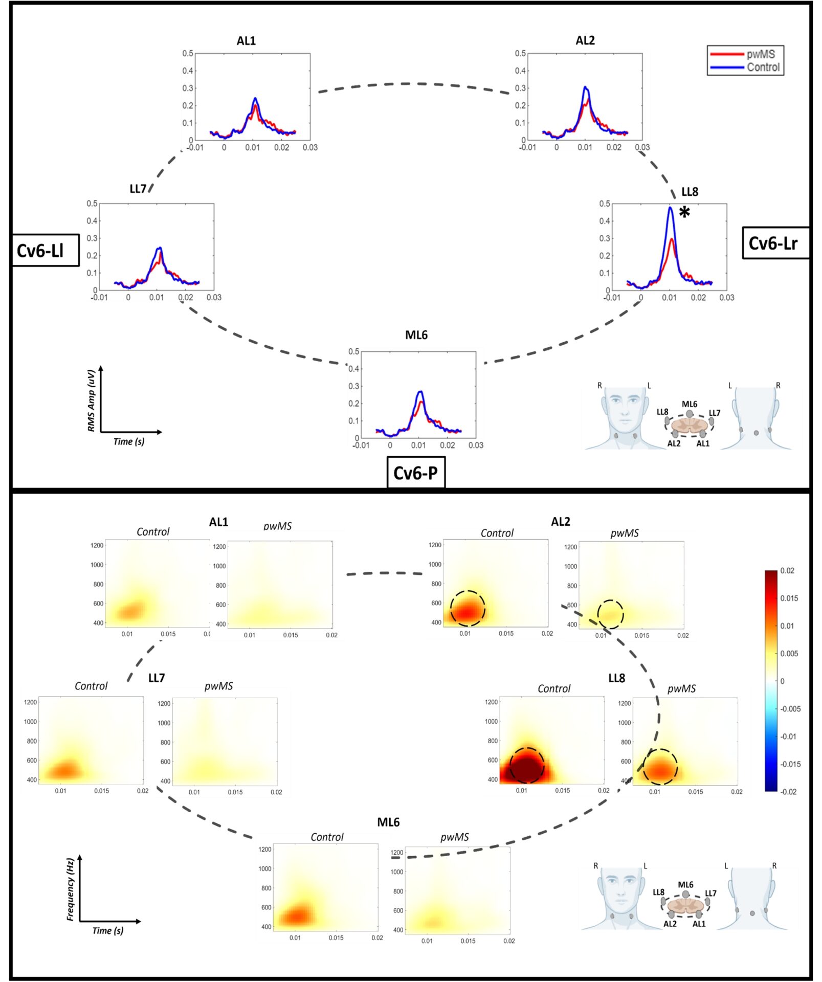
A highly lateralised (p < 0.05) evoked HFOs were observed at lower cervical levels, with the maximum response observed at the LL8 electrode (C6 vertebral level (Cv6): right-lateral channel) for both groups [pwMS: 0.26 ± 0.11, controls: 0.4 ± 0.2]. The observed Peakamp at the LL8 electrode was significantly lower in pwMS (p = 0.021). No significant differences were observed for onset or peak latency.
During the 8-15 ms post-stimulation period, both groups demonstrated broadband activity between 400-600 Hz at lower cervical levels. However, this activity was significantly reduced in the pwMS.
Discussion:
This first-of-its-kind study reveals specific disease-relevant alterations in pwMS, highlighting the potential of HD-ESG-based investigations towards a viable marker of spinal dysfunction. The reduction in peak HFO amplitude likely reflects temporal dispersion due to demyelination in MS[6], indicative of impaired cervical spinal integrity. A similar decrease in HFO amplitude has previously been reported at the somatosensory cortex in pwMS[6]. Our findings indicate that these alterations are detectable even at the cervical spinal level, suggesting that HFO-alterations may represent a signature of network-level dysfunction.