Background: Traumatic injuries are known to modulate the neuromuscular system and adapt to the feedback processes interrupted, altered or removed by injury (1). Here, we present a case-study of a 26-year-old male (height 178.4cm, mass 67.3kg) who was bitten by a shark on his left lateral thigh whilst surfing. To restore function, a free muscle flap from the latissimus dorsi was inset to the remaining rectus femoris (RF). Following intensive rehabilitation, this surfer returned to surfing at a high level. Using transcranial magnetic stimulation (TMS), we explored how the supraspinal pathways to the lower limbs were affected.
Methods: The protocols were approved by the institution’s Human Research Ethics Committee (16/133). Surface electromyography (sEMG) recordings were captured bilaterally from the vastus lateralis (VL), vastus medialis (VM) and RF using bipolar electrodes. On the left, or affected limb (AL), sEMG could not be placed in a similar position to the right, or unaffected limb (UAL), for both VL and RF. The VL sensor was therefore placed over the free muscle flap for the injured RF (RF-Lat), while RF sensor was placed over the proximal, remaining portion of RF (RF-Prox). TMS-induced motor evoked potentials (MEP) were generated at 120% of the active motor threshold, with the participant receiving 20 stimuli in both a standing and supine condition. During the supine condition, sEMG activity was matched to standing for VM of the AL. Waveforms were normalised to their root mean square amplitude. MEP area and cross-correlation lag were compared between conditions and limbs for each muscle, with the waveform differences compared using Statistical Parametric Mapping (SPM; 2).
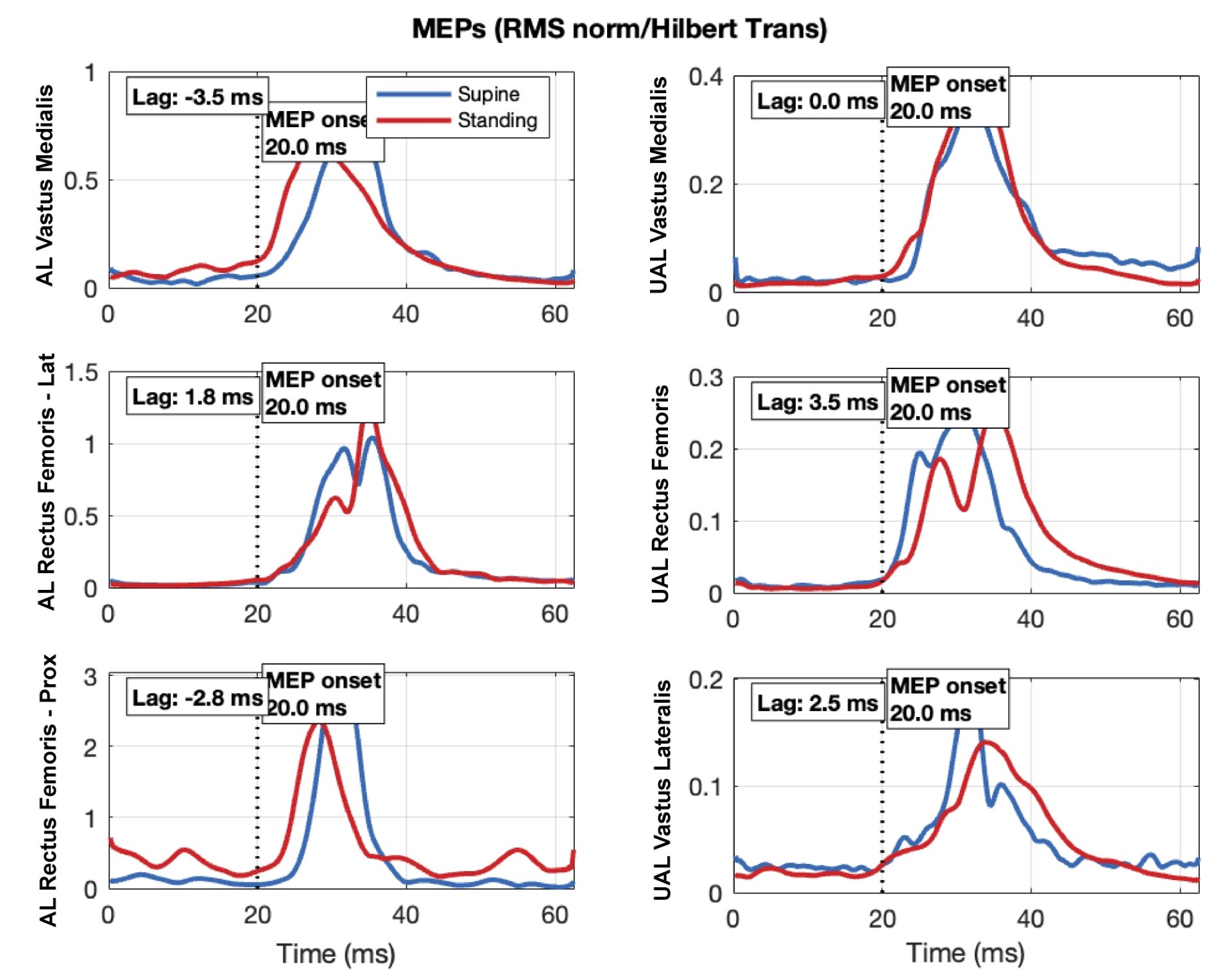
Results: The transition from supine to standing significantly increased MEP area (p < 0.05), including for the VM where the matched sEMG activity would equalise the state of the motoneuron pool. In the AL, cross-correlation lag was -3.5 and -2.8 ms during standing in the VM and RF-Prox, respectively (Figure 1). This was supported by the SPM analyses, which indicated VM and RF-Prox had an earlier onset in the AL when comparing standing to supine. Importantly, the RF-Lat demonstrated no change in MEP onset with the transition to standing, following similar results to the UAL muscles.
Conclusion: The results provide a unique appreciation for how the neuromuscular system adapts to postural challenges following traumatic injury. The earlier MEP onset of the AL VM and RF-Prox suggest a different innervation pattern to the RF-Lat and UAL muscles, which may be explained by two potential mechanisms. This shift in MEP timing may be driven by an increase in excitability of the motoneuron pool for VM and RF-Prox, which is plausible considering that for VM this was matched between conditions to equalise the state of the motoneuron pool. Alternatively, this may be due to changes in neuromuscular state that preferentially increased the excitability of direct corticomotoneuronal cells during standing. In both cases, this may facilitate faster signal transmission to the knee of the AL during dynamic tasks to account for its functional limitations in strength.