
Astrocytes are necessary to the maintenance of the blood-brain barrier (BBB; Janzer & Raff, 1987). However, the mechanism by which they induce the BBB phenotype is still unclear. The astrocytes and the cerebral endothelial cells are separated by two basement membranes, which are likely to be involved in the interactions between the two cell types. Laminin, a basement membrane constituent, has been reported to interact with several protein complexes present on the astrocytes and the endothelial cells. One of these complexes is the dystrophin glycoprotein complex (Khurana et al. 1995), but its specific cellular localisation is unclear (Knuesel et al. 2000; Ueda et al. 2000). Previous studies have also shown that the brain and spinal cord show different susceptibilities to endothelial barrier breakdown, but the reasons underlying this disparity are unknown (Schnell et al. 1999). In this in vivo study, we used immunohistochemistry and confocal microscopy to establish which cells express the various components of the dystrophin complex in naive mouse brains (striatum, n=3) and spinal cords (thoracic, n=3). The expression of the proteins of the dystrophin complex was also quantified in these two compartments and correlated to susceptibility. The brains and spinal cords were isolated from mice killed humanely by an overdose of sodium pentobarbital. The principal findings of this study are: (i) endothelial cells and astrocytes do not express identical dystrophin complex proteins. Astrocyte express dystrophin, dystroglycans and dystrobrevins while endothelial cells only express utrophin; (ii) there is a clear difference in the expression pattern of laminin and the dystrophin complex proteins in the brain and spinal cord and in the grey and white matter. Expression of these proteins was consistently higher in the brain white matter compared with the other compartments (see Fig. 1, Student’s t test). This study has shown that astrocytes and endothelial cells express distinct dystrophin complexes, suggesting that these complexes exert different functions. The utrophin ligands remain to be identified. The brain and the spinal cord basement membrane are quantitatively different but these differences are only small and are unlikely to wholly explain why the brain and the spinal cord show different susceptibilities to injury. However, the vascular basement membranes of the brain grey and white matters are significantly different which may partly explain the different susceptibilities of these two regions to inflammation and injury.
King's College London (2005) J Physiol 565P, PC144
Communications: Expression of the Dystrophin-Glycoprotein Complex and its Ligands at the Blood-Brain Barrier
Bernardes-Silva, Martine ; Forse, Penny A; Perry, V Hugh;
1. CNS Inflammation Group, University of Southampton, Southampton, United Kingdom.
View other abstracts by:
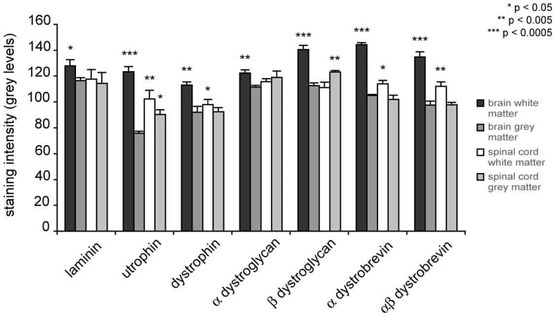
Figure 1. Quantification of laminin and the dystrophin complex proteins immunostaining in the vascular basement membranes of naive mouse brain and spinal cord grey and white matters.
Where applicable, experiments conform with Society ethical requirements.