
Hyperammonemia-induced increases in brain lactate have been studied mainly with protocols designed to mimic hepatic encephalopathy, but it has been suggested that there is a physiological flux of ammonium from neurons to astrocytes as part of glutamate-glutamine shuttling (1). In vitro, ammonium activates enzymes of glycolysis and inhibits the TCA cycle, and so tends to increase lactate production. Also, NH4+ is avidly taken up by astrocytes (2,3) and there is evidence for a metabolic flux through lactate in the brain during normal activity (4). Minimum conditions for a possible role of ammonium in regulating lactate production are that ammonium-induced increases in lactate be rapid and reversible. We have used localized 1H NMR spectroscopy at 7 Tesla to monitor changes in the concentrations of brain metabolites. Rats were anæsthetised with 5% isofluorane and maintained with mechanical ventilation with 1–1.5% isofluorane in air enriched with O2 to 35%. A 4 x 4 x 4 mm3 voxel was centred on the striatum, the echo time was 136 ms, and spectra were acquired over 3 min 12 s. We measured the areas of peaks corresponding to choline compounds (tCho), creatine /creatineP (tCr), N-acetyl aspartate (NAA) and lactate. In Fig. 1, when 1M NaCl was infused through a femoral vein over 4 min at a rate that gave a total quantity of 2.5 mmol/(kg body wt) the lactate signal did not change. A second infusion, identical except that NaCl was replaced by NH4Cl, caused lactate to increase 5-fold. All but about 2.4% of the ammonium had been cleared from the blood by 1 min after the end of the infusion. On average, control infusions of NaCl caused no change in the signals for lactate or other metabolites except for tCr (–3.07%, s.e.m. = 0.81%, 5 infusions in 5 rats, P < 0.02, all Ps by Student's two-tailed t test). With NH4Cl, the lactate signal started to increase during the infusion and reached a peak 3.10 ± 0.35 times baseline (P < 0.0001) at 13.2 ± 2.1 min after the infusion (9 infusions in 5 rats). It then recovered halfway to baseline by 31.2 ± 5.7 min after the infusion. tCr and tCho signals were unchanged. The NAA signal increased by 3.52 ± 0.68% (P < 0.0006). In parallel experiments, changes in cerebral blood flow were measured by inverting the magnetization of water protons in the carotid arteries and detecting the ensuing signal changes in the brain (5). After ammonium infusion, cerebral blood flow increased by a factor of 2.15 ± 0.16 (5 infusions in 3 rats, P = 0.0002), suggesting that the lactate increase was not caused by hypoxia. Ammonium-induced increases in lactate signal could be observed at least three times in one experiment.
University College London 2006 (2006) Proc Physiol Soc 3, C19
Research Symposium: Monitoring rat brain lactate concentration in vivo by NMR shows rapid, reversible, increases induced by brief infusions of ammonium
Peggy Provent1, Nils Kickler1, Régine Farion1, Emmanuel L Barbier1, Païkan Marcaggi2, Christoph Segebarth1, Jonathan A Coles1
1. INSERM U594, Université Joseph Fourier, Grenoble, France. 2. Physiology Department, University College London, London, United Kingdom.
View other abstracts by:
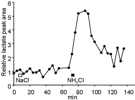
Figure 1. Time course of changes in the area of the lactate peak in an illustrative experiment. Infusion of NaCl over 4 min caused no change but NH4Cl did. Data acquisition lasting 3 min 12 s was started simultaneously with the beginning of the NH4Cl infusion and this point already showed an increase.
Where applicable, experiments conform with Society ethical requirements.