
Age-related declines in cellular structure and function can be initiated or accelerated by physical inactivity. Ageing is associated with aberrant nuclear morphology, architecture and translation of forces to biochemical signals (mechanotransduction); this project focuses on whether, in skeletal muscle, these changes are influenced by physical activity status. Nuclear morphology and the distribution of nuclear envelope proteins important for nuclear architecture and mechanotransduction (SUN1 and LEM2) were studied in isolated human muscle fibres from individuals of different ages and physical activity statuses. So far, the data indicate that in humans, changes in myonuclear morphology occur in response to exercise regardless of age, indicated by a 25-30% reduction in myonuclear aspect ratio in young and older exercise-trained individuals compared to inactive young and older counterparts. No differences in the distribution of proteins SUN1 or LEM2 were revealed through super-resolution microscopy. Exercise therefore appears to influence nuclear morphology in a manner unaffected by age; alterations to nuclear envelope proteins other than SUN1 and LEM2 may be involved in this process. Future research will be focused on analysing myonuclear morphology in 3D, the distribution of nuclear envelope proteins Lamin A and Nesprin-1 and muscle-specific cytoskeletal protein Desmin. Whether alterations to myonuclear mechanics influence gene transcription will also be assessed through cell microharpooning and RNA sequencing.
Future Physiology 2019 (Liverpool, UK) (2019) Proc Physiol Soc 45, C08
Oral Communications: The missing LINC to human healthy ageing: myonuclear architecture and mechanics in old age
E. Battey1,2, S. Harridge1, R. Pollock1, N. Lazarus1, M. Stroud2, J. Ochala1
1. Centre for Human & Applied Physiological Sciences, King's College London, London, United Kingdom. 2. British Heart Foundation Centre of Excellence, School of Cardiovascular Medicine and Sciences, King's College London, London, United Kingdom.
View other abstracts by:
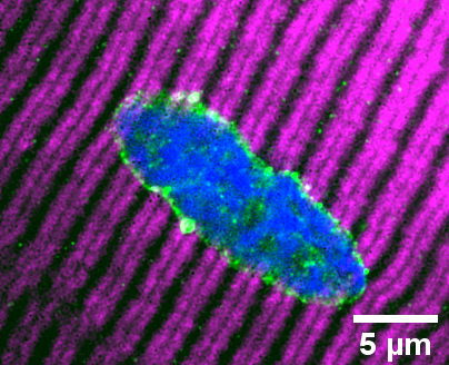
Myonucleus of an isolated skeletal muscle fibre extracted from a vastus lateralis biopsy of a young inactive individual. Myonuclei in these individuals, and in older hip fracture patients (model of disuse), are elongated, which contrasts rounder myonuclei in active younger and older counterparts. Fibres stained to visualise Myosin heavy chain 7 (magenta), nuclear envelope/LINC complex protein SUN1 (green) and DNA (blue).
Where applicable, experiments conform with Society ethical requirements.