
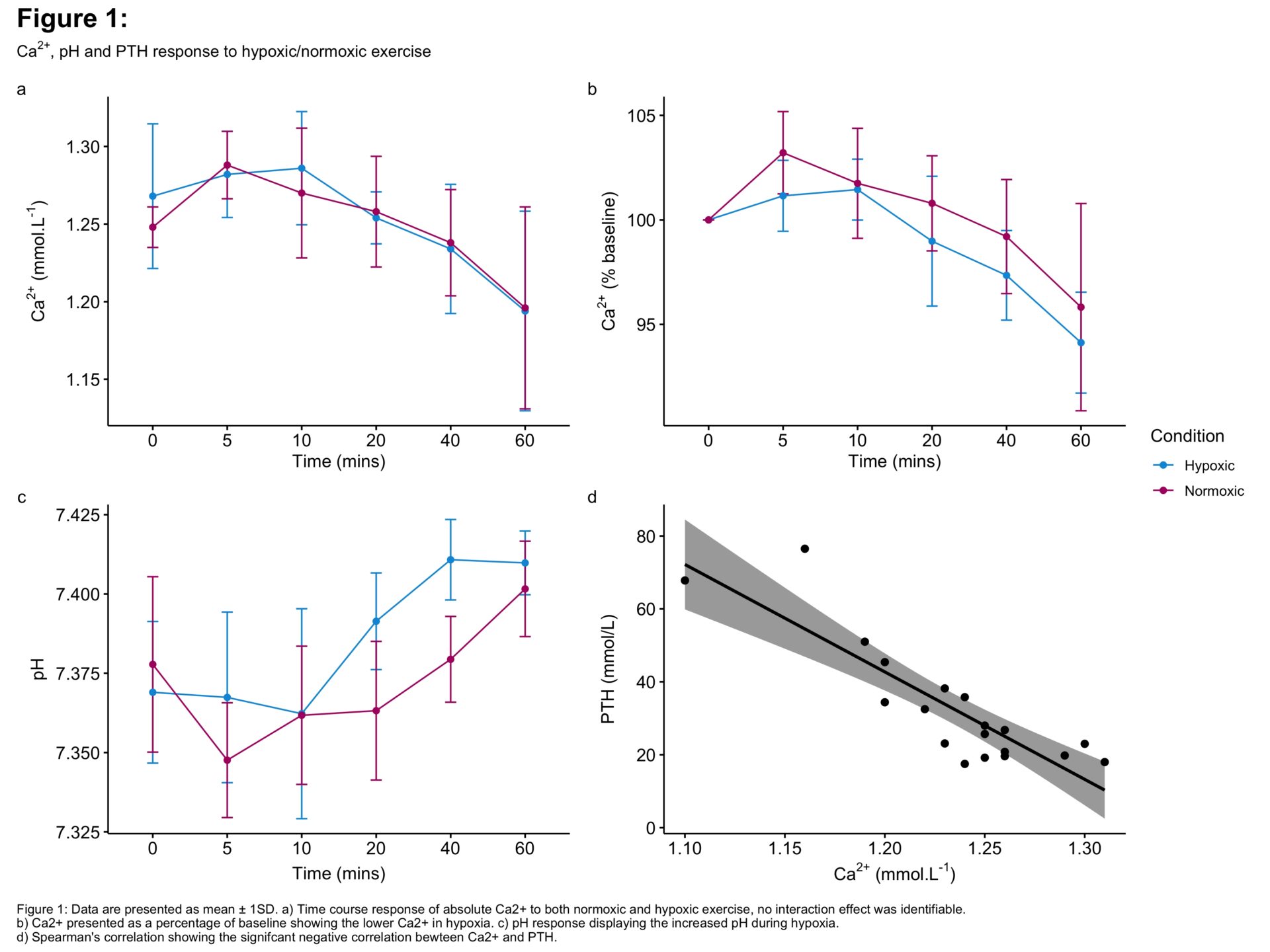
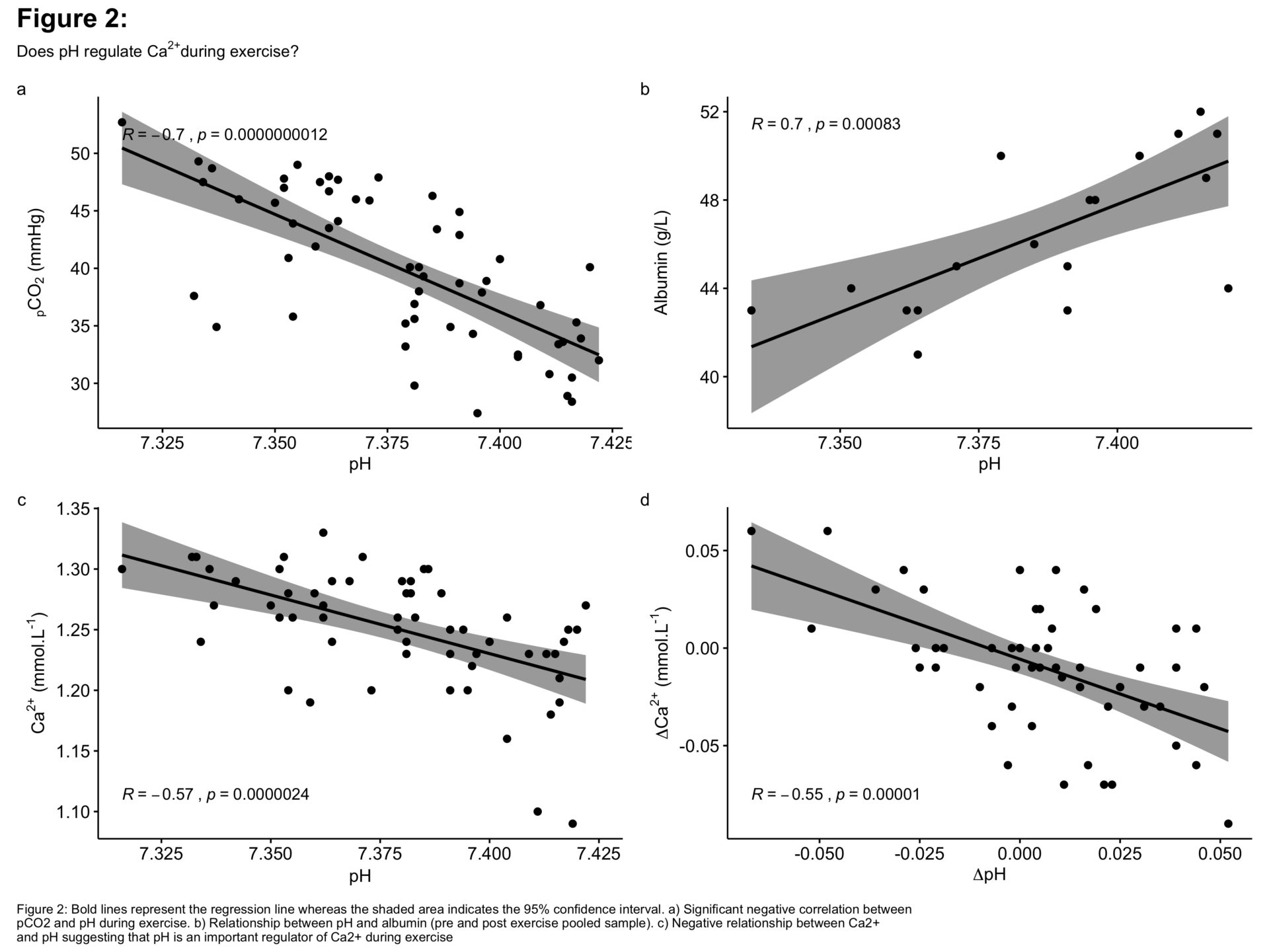
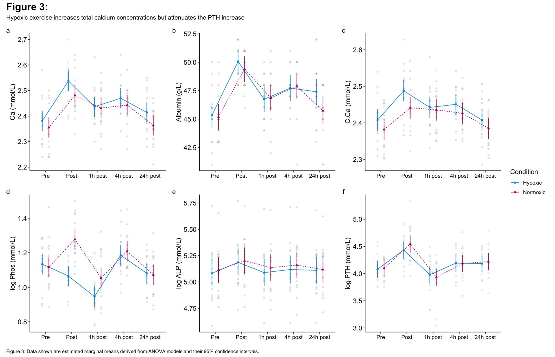
Several investigations show exercise induces a decrease of ionised calcium which may be responsible for increased bone breakdown following acute exercise (1-3). While the general mechanism of decreased Ca2+stimulating PTH and subsequent bone resorption has been comprehensively detailed (1-2), what causes Ca2+ to decrease remains unknown. We suggest it may be the result of the complex acid-base changes seen during exercise (4) as Ca2+ is heavily influenced by pH. We investigated whether pH alterations, achieved by use of exercise and hypoxia, influenced calcium metabolism. Sixteen healthy physically active males volunteered for our study ([mean ± 1 SD] age 24 ± 5 years, height 1.80 ± 0.08m, body mass ± 78.5 ± 12.6kg), approved by the University’s Research Ethics Committee and in accordance with the Declaration of Helsinki. Participants were randomly assigned in a counter-balanced manner to perform two V̇O2max tests on a cycle ergometer in either normoxia (21% FiO2) or hypoxia (13% FiO2), one week apart. Participants then completed 60 minutes of cycle exercise at 70% of their respective V̇O2max/peak in both conditions. Venous blood was collected via venepuncture: pre-exercise; immediately post- exercise; and 1h, 4h and 24h post exercise, to measure markers of calcium metabolism. A sub-sample of participants (n = 5) provided additional blood samples during the exercise trials (see Figure 1a) for analysis of Ca2+ and pH. Data were analysed using two-way repeated measures ANOVAs and regression. We observed that exercise caused a transient increase in Ca2+ at the onset of exercise and later decrease until cessation (Figure 1a). Ca2+ was positively correlated with PTH (Figure 1d: R = -0.82, p < .001) further supporting previous observations. Although hypoxic exercise reduced relative Ca2+ to a greater degree, and despite pH being higher during hypoxia (Figure 1c: Condition*Time p = .01), the interaction of time and condition was non-significant. The respiratory alkalosis during hypoxia appeared to reduce competition for albumin binding positions, demonstrated by the positive correlation between pH and albumin (Figure 2b: R = 0.7, p < .001). We speculate that a greater sample size may observe a difference between conditions. The higher total calcium concentration following hypoxic exercise (Figure 3c; MD = 0.04±0.01, p < .001) and the negative correlation between Ca2+ and pH (Figure 2c-d: R = -0.57, p < 0.0001) support that Ca2+ is bound to albumin during respiratory alkalosis. Hypoxia attenuated the phosphate response to exercise, demonstrated at the post exercise time point (Figure 3d: MD = -0.21±0.02, p < .001), which may be explained by hypocapnia. The reduced CO2 increased pH (Figure 2a), known to stimulate the glycolytic system and the production of phosphofructokinase (PFK). PFK enhances sugar phosphate production and subsequently stimulates phosphate entry to muscle (5). It is possible that the decrease in phosphate may have been compounded by the hypoxic-induced transcription of fibroblast growth factor 23 (FGF23). FGF23 is known to reduce serum phosphate concentrations and negatively regulate PTH, possibly explaining the lower PTH response post hypoxic exercise (Figure 3f; MD = -0.11 ± 0.05, p < .05).
Future Physiology 2020 (Virutal) (2020) Proc Physiol Soc 46, PC0029
Poster Communications: The Effect of Acute Hypoxic Exercise on Calcium Metabolism
Scott Hannah1, Conor McClean1, Sonyia McFadden2, Andrea McNeilly1
1 Ulster University, Sport and Exercise Science Research Institute, Jordanstown, Belfast, United Kingdom 2 Ulster University, Institute of Nursing and Health Research, Jordanstown, Belfast, United Kingdom
View other abstracts by:
Where applicable, experiments conform with Society ethical requirements.