Introduction: Ageing is typified by a decline in maximal oxygen uptake (VO2max) (Conley et al., 2000a; Conley et al., 2000b), which can limit physical function and daily life activities. This decline in VO2max is related to a lower skeletal muscle mitochondrial function and loss of muscle mass (Conley et al., 2000a; Conley et al., 2000b). However, data in humans are inconsistent, with many studies showing no difference in mitochondrial function between young and old or middle-aged groups (Rasmussen et al., 2003). Most human studies have relied on assessments of maximal mitochondrial function (i.e. enzyme activities or phosphocreatine recovery kinetics), which may conceal subtle changes in mitochondrial ultrastructure and function occurring prior to an overt reduction in maximal mitochondrial oxygen consumption.
Objectives: To determine aerobic exercise capacity, mitochondrial respiration, density, and ultrastructure in late middle-aged and younger adults, and systematically assess the interrelationships between VO2max and muscle mitochondrial measures.
Methods: 12 healthy young (27 ± 5 years, 8 males) and 10 late middle-aged adults (55 ± 6 years, 5 males) were recruited. All participants performed a maximal ramp incremental test on a cycle ergometer to determine VO2max. On a separate day, muscle biopsies were obtained from the vastus lateralis muscle. Mitochondrial respiration was assessed in permeabilized fibres using high-resolution respirometry. Transmission electron microscopy was to study mitochondrial density and ultrastructure. Succinate dehydrogenase (SDH) activity was assessed via histochemistry in sections.
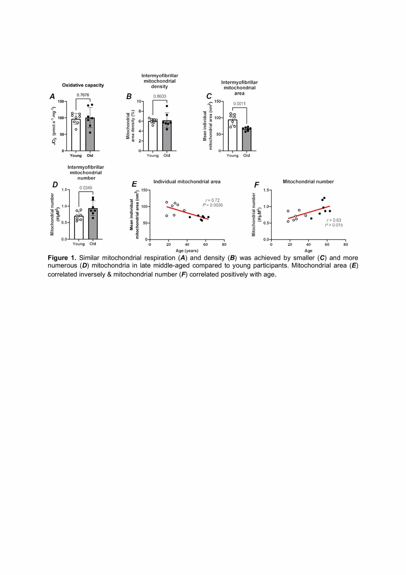
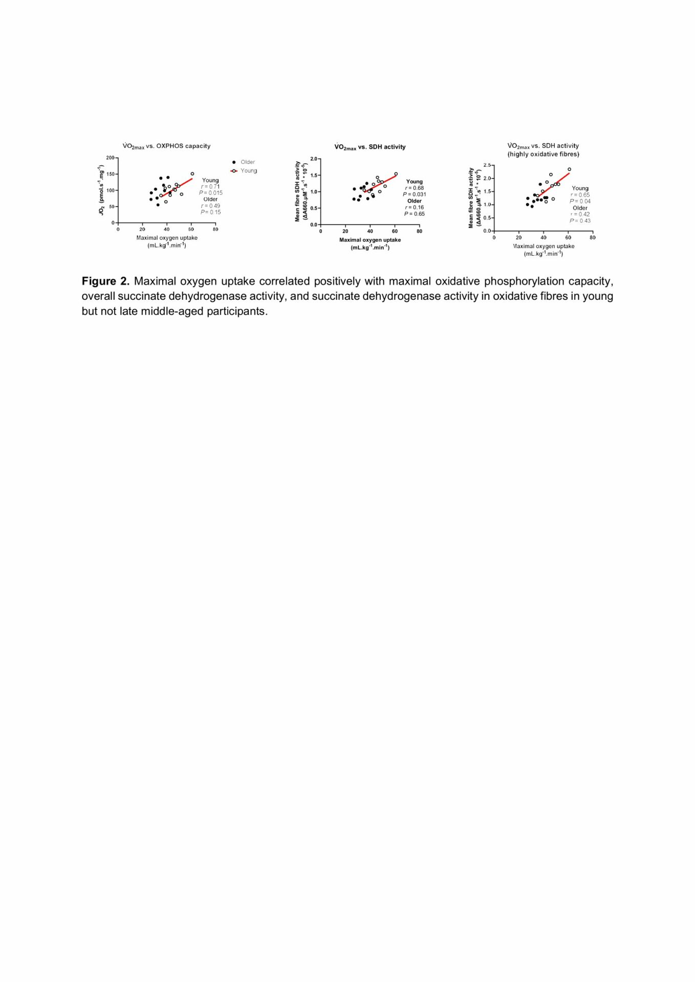
Results: VO2max was lower in the late middle-aged compared to the younger group (34±5 vs. 45±7 mL.kg-1.min-1, P<0.001). Despite this, maximal oxidative phosphorylation capacity (old = 99±27 vs. young = 99±17 pmol O2.s-1.mg-1, P=0.95) and mitochondrial area density (old = 6.2±1.5 vs. young = 6.0±0.5%, P = 0.86) did not differ between groups. SDH activity was lower in old versus young subjects (old = 0.97±0.18 vs. young = 1.18±0.20 *10-5 ΔA660.µM-1.s-1, P=0.02), an effect that appeared to be confined to the most oxidative fibres (old = 1.23±0.22 vs. young = 1.65±0.39 *10-5 ΔA660.µM-1.s-1, P =0.008) and not the least oxidative fibres (old = 0.71±0.22 vs. young = 0.75±0.13 *10-5 ΔA660.µM-1.s-1, P=0.69). Late middle-aged participants displayed smaller (old = 66±5 vs. young = 95±17 nm2, P=0.001), but more numerous (old = 0.71±0.13 vs. young = 0.94±0.22 mitochondria.µm-2, P=0.03) mitochondria when compared to younger participants. The area of individual mitochondria correlated negatively, and the number of mitochondria per unit area of muscle correlated positively with age, respectively (Figure 1). Various indices of mitochondrial function were correlated with VO2max in the younger but not the late middle-aged group (Figure 2).
Conclusion: These data demonstrate that late middle-aged individuals had smaller, more numerous mitochondria for the same mitochondrial density and maximal respiration. The lack of correlations between mitochondrial measures and VO2max in this group suggests that VO2max is more likely constrained by O2 delivery-related processes in late middle-aged humans. The extent to which these mitochondrial structural alterations predispose skeletal muscle to ageing remains to be determined.