
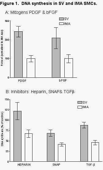
Saphenous vein (SV) bypass grafts have poor long-term patency compared to internal mammary artery (IMA) grafts [1]. Smooth muscle cell (SMC) proliferation is a major component of intimal hyperplasia and vein graft disease. This study indicates that intrinsic differences in SMC proliferative capacity may contribute to the different patencies of arterial and venous grafts. With informed consent and local research ethics committee approval, matched SV and IMA SMCs were isolated by explant culture from surplus graft tissue within six individual patients undergoing coronary artery bypass surgery. In the presence of 20% fetal calf serum (FCS), venous and arterial explant outgrowth rates were compared and population growth rates of extracted SMC at passage 4 were analysed over 8-10 days using a resazurin dye method. DNA synthesis experiments using 3H-thymidine incorporation were performed on cells rendered quiescent in low serum before stimulation with platelet derived growth factor (PDGF, 30ng/ml) or basic fibroblast growth factor (bFGF, 20ng/ml). The effects of heparin, transforming growth factor β (TGF-β) and increased nitric oxide produced by the nitric oxide donor S-nitroso-N-acetylpenicillamine (SNAP) were assessed on DNA synthesis induction by 5% FCS. Data are presented as mean ± standard error of the mean. One-way ANOVA was used to determine differences between groups and treatments with a P value of <0.05 considered significant. Although SMC proliferation rates varied considerably between patients, SV SMC populations increased significantly faster than IMA measured as outgrowth from explant (not shown); increase in cell number over 8 days (7860 ± 1300 vs 4514 ± 930) and DNA synthesis induction with PDGF (1143 ± 89.9 vs 594 ± 96.2) and bFGF (407 ± 138 vs 195 ± 66). In addition, heparin stimulated DNA synthesis in SV SMC (121.6 ± 20.41% of control) but significantly inhibited IMA SMC (67.3 ± 9.2%). SV SMCs were also less sensitive than IMA SMCs to the inhibitory effects of TGF-β (88.0 ± 6.8% vs 45.8 ± 5.5% of control) and SNAP (68.7 ± 8.0% vs 41.7 ± 4.1% of control). Using paired SV and IMA SMC cultures we have demonstrated that, despite large variations between individuals, venous SMCs have a significant growth advantage over arterial counterparts. This is due to enhanced responses to growth factors and reduced sensitivity to endothelium-derived and other inhibitors of proliferation and implies an extra level of proliferation control in arterial SMCs.
University of Oxford (2005) J Physiol 568P, PC59
Poster Communications: Differential responses to growth factors and inhibitors in arterial and venous smooth muscle cells derived from bypass graft tissue
Lowe, Robert; Cross, Nicola; Browning, Paul;
1. Research, The Cardiothoracic Centre, Liverpool NHS Trust, Liverpool, United Kingdom.
View other abstracts by:
Where applicable, experiments conform with Society ethical requirements.