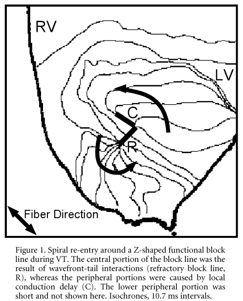
Spiral re-entry plays an important role in the initiation and maintenance of ventricular tachyarrhythmias (Pertsov et al. 1993; Fast & Kleber, 1997). However, the precise dynamics of spiral wave propagation in the heart, which determines the behaviour of the activity, are not fully understood. In the present study, we investigated dynamics and pharmacological modulation of spiral re-entry during ventricular tachycardia in a two-dimensional anisotropic subepicardial sheet of ventricular myocardium by optical mapping. Rabbits were killed humanely with an overdose of sodium pentobarbital (n = 7). A Langendorff-perfused epicardial layer (~1 mm thick) of rabbit ventricle was made by cryoablation of the endocardial side of the left ventricle. The heart was stained with a potentiometric dye, di-4-ANEPPS, and fluorescence images of the left ventricle were obtained via a high-speed video camera with temporal and spatial resolution of 1.3 ms and 0.1 mm, respectively. In 4 of 7 cases, ventricular tachycardia (VT) induced by cross-field stimulation showed a single circular re-entry around a Z-shaped functional block line (Fig. 1).
The Z-shaped block line was composed of two portions in which the underlying mechanisms were different: the central portion of the block line was created by meandering of the spiral tip along its own refractory tail, and the two peripheral limbs were caused by pronounced local conduction delay at the pivot points. The peripheral block lines were always oriented parallel to the epicardial fibre direction. Double-humped action potentials were recorded at the central block line. Na+ channel blockade by disopyramide or pilsicainide enhanced local conduction delay at the pivot points leading to an extension of the peripheral block lines. Around the pivot points, action potentials were widened with steps in the depolarization phase, whereas after making a turn around the pivot points, action potentials were relatively short, giving rise to an excitable gap. These results suggest that the dynamics of spiral re-entry in the two-dimensional ventricular myocardium are mainly determined by (1) tissue anisotoropy, (2) wavefront curvature and (3) the wavefront-tail interaction.
All procedures accord with current National guidelines.