
Lymphoedema distichiasis (LD) is a dominant inherited primary lymphoedema with onset of bilateral lower limb swelling around puberty, in association with aberrant eye lashes from the meibomian glands (distichiasis) and venous abnormalities. Causative mutations have been described in the FOXC2 gene. The mutation is presumably present in all lymphatics, yet only the legs swell. We therefore tested the hypothesis that leg dependency adversely affects initial lymphatic filling in those with LD compared with normals. Of 15 volunteers, 10 aged 21-61y had a FOXC2 mutation, leg lymphoedema and distichiasis; 5 were family members (23-57y) without FOXC2 mutation, lymphoedema or distichiasis. Ethics committee approval and informed, written consent were obtained. The dermal lymphatic plexus of the foot was imaged first when dependent and then at heart level after 60-80 min supine. 5 μl fluorescein isothiocyanate dextran (150 kDa, 25%w/v) was injected intradermally and its uptake into the initial lymphatics monitored by fluorescence videomicroscopy for 45 min. A lymphatic map of vessels was analysed stereologically for total length of filled lymphatics (LL), maximum lymphatic length density (LDmax), maximum dye spread and sector filling (ref 1). There were no significant quantitative differences between the initial lymphatic networks visualised in the dependent and supine positions in control feet (Table 1, n = 5, mean ± s.d.). Microlymphatic filling in the dependent position in LD was significantly reduced when compared with the supine position (Table 2, n = 10). We conclude that during dependency, flow into the initial lymphatic network in LD patients is impaired. A possible cause of reduced filling is lymph reflux due to collecting lymphatic valve failure. There may also be a role for abnormal recruitment of mural cells around initial lymphatic vessels (ref 2).
King's College London (2005) J Physiol 565P, C131
Communications: Impaired filling of initial lymphatics in lymphoedema distichiasis during dependency
Mellor, R H; Tate, N ; Stanton, A WB; Brice, G ; Sholto-Douglas-Vernon, C ; Mansour, S ; Child, A H; Burnand, K G; Jeffery, S ; Levick, J R; Mortimer, P S;
1. Lymphoedema Research Group, St George's Hospital Medical School & *St Thomas' Hospital, London, United Kingdom.
View other abstracts by:

Table 1: Lymphatic network measurements in controls

Table 2: Lymphatic network measurements in LD group
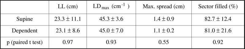
Table 1: Lymphatic network measurements in controls
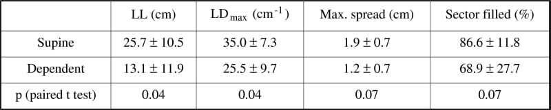
Table 2: Lymphatic network measurements in LD group
Where applicable, experiments conform with Society ethical requirements.