Muscle spindles are mechanosensors crucial for proprioception and skeletal alignment. Previous studies have shown a column of more than 20 muscle spindles within the rodent deep masseter muscle, including rats and mice, suggesting a specialised role in sensory feedback during jaw movement. Mitochondrial defects can cause malocclusion, a congenital misalignment of the jaws. Interestingly, we found that muscle spindle sensory terminals are densely packed with mitochondria, occupying more than 50% of the terminal volume. To investigate how the spindle column arises and is affected by mitochondrial defects, we are determining its precise location and structure throughout development, then in a mouse model of malocclusion.
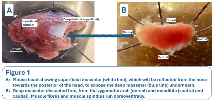
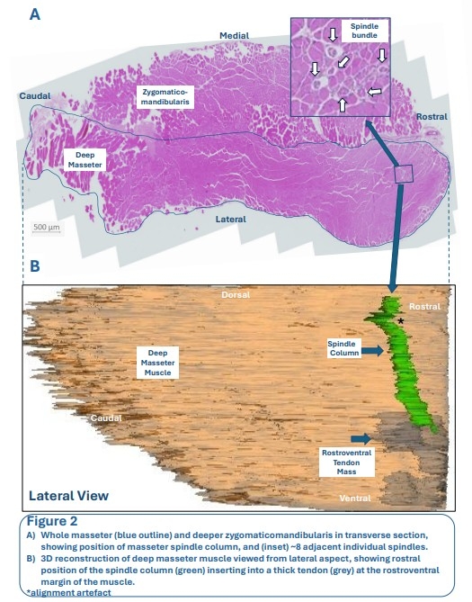
This initial study aims to develop a technique for creating a 3D model of the spindle column in adult mouse muscle. The right deep masseter muscle was dissected, fixed in 4% paraformaldehyde overnight, before dehydration and wax embedding. The sample was serially sectioned transversely at 5 μm thickness, then stained with haematoxylin and eosin. Sections were scanned, and images were manually aligned using GIMP image processing software. Reconstruct software was used to trace manually the muscle and muscle spindle outlines within each section, then compiled to provide the 3D visualisation of their spatial distribution.
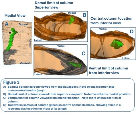
Preliminary findings confirmed the spindle column’s presence near the masseter’s anteromedial margin and provided the first comprehensive histological map. Future work will determine the column’s location throughout development, and whether malfunctioning mitochondria disrupt its alignment and contribute to malocclusion.