PKC isoforms play a major role in ischaemic preconditioning (IPC) of the human heart (Speechly-Dick et al. 1995). Using specific PKC isoforms inhibitors we aim to investigate which PKC isoforms are involved in IPC of the human myocardium and to determine their relation to mitoKATP channels.
The study was approved by the local ethical committee and was conducted with the consent of patients. Right atrial muscles obtained from patients undergoing elective cardiac surgery were equilibrated according to our model (Zhang et al. 2000). Tissue sections were randomized to receive any of the following protocols (n = 6 per group): (1) aerobic control, (2) simulated ischemia for 90 min /reoxygenation for 120 min (SI/R), (3) IPC using five min SI followed by five min reoxygenation, (4) the addition of PKC isoforms inhibitors 10 min before and 10 min during IPC. PKC isoforms inhibitors investigated were V1-2 peptide [10 µM], GO6976 [100 nM], Rottlerin [100 µM] and LY333531 [100 nM] for PKC  , PKC α, PKC δ and PKC β respectively.To investigate the relation of PKC isoforms to mitoKATP channels, PKC inhibitors found to be involved in IPC were added 10 min before and 10 min during preconditioning by Diazoxide (DXZ)[100 µM] in a second experiment (n = 6 per group). CK leakage and 3-[4,5-dimethylthiazol-2-yl]-2,5-diphenyltetiazolium bromide (MTT) cell viability were measured. All the values were expressed as mean ± S.E.M., *P < 0.05 paired t test). Phosphorylation of PKC isoforms following activation of the sample by either DXZ or IPC was detected using western blotting and then analysed using Scion image software.
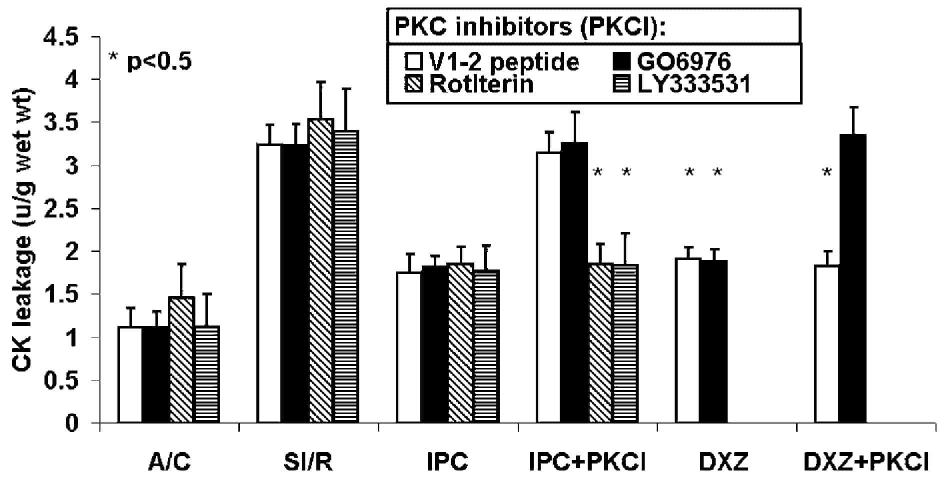
In the Figure, PKC and PKC α inhibitors blocked IPC whereas PKC δ and PKC β inhibitor did not. The protection elicited by mitoKATP channels opening with DXZ was blocked by the inhibition by PKC α isoform but not by inhibition of PKC isoform. The MTT results were a mirror image of the CK results. In addition DXZ caused increased phosphorylation of PKC α similar to IPC but failed to cause significant increase of PKC phosphorylation.
PKC α and PKC are involved in IPC of the human myocardium with PKC upstream and PKC α downstream of mitoKATP channels.
We acknowledge the support of D.A. Fowler and Prof M. Galinanes.