
Immature, non-contractile myocytes morphologically similar to the intestitial cells of Cajal, have been found resident in the vasculature1. It is thought that these cells are synthetic vascular myocytes and play a major role in vascular remodelling2. This work aimed to determine the quantitative differences in transcription of smooth muscle cell and interstitial cell marker genes between immature and mature vascular myocytes. Genes investigated were: smooth muscle (SM) α-actin, β-actin, myosin light chain kinase (MLCK), desmin, smooth muscle myosin heavy chain (SM-MHC) isoforms SM1 and SM2 (smooth muscle marker genes) and c-kit and vimentin (markers of interstitial cells). Investigations were carried out on freshly isolated myocytes from mesenteric arteries of guinea-pigs which were killed in accordance with national guidelines for humane killing of experimental animals (Schedule 1). Single mature and immature myocytes were obtained by enzymatic digestion and collected using a wide-bore pipette mounted on a micromanipulator. Cells were prepared for qRT-PCR using the TaqMan PreAmp Cells-to-Ct Kit (Applied Biosystems). This involved cell lysis, DNA removal, reverse transcription of RNA into cDNA and gene-specific preamplification using custom assays followed by analysis in a qRT-PCR analyser. Custom TaqMan gene expression assays were designed for each gene using available guinea-pig sequences and sequences determined by homology. CT values were compared directly between cell phenotypes and fold differences (ratio of mRNA quantity in mature cells versus immature cells) for each target gene were tested for statistical significance using one sample Student’s t-test (p=0.05). The results are shown in Table 1. C-kit mRNA was not detected in either mature or immature myocytes (n=13, N=4), but was present in the wall of the small intestine (n=3, N=1), confirming the validity of the assay. These data confirm common cell lineage for immature and mature vascular myocytes and identify subtle differences in gene expression pattern between them, which hint at different physiological roles of these two phenotypes.
King's College London (2008) Proc Physiol Soc 13, PC5
Poster Communications: Quantitative RT-PCR analysis of smooth muscle markers between mature and immature vascular myocytes
J. Lynch1, A. Collins1, V. Pucovský1
1. Queen's University Belfast, Belfast, United Kingdom.
View other abstracts by:
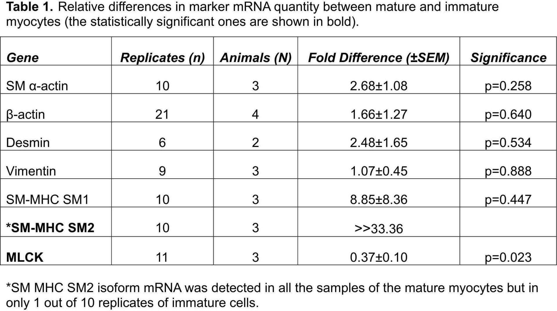
Table 1. Relative differences in marker mRNA quantity between mature and immature myocytes (the statistically significant ones are shown in bold).
Where applicable, experiments conform with Society ethical requirements.