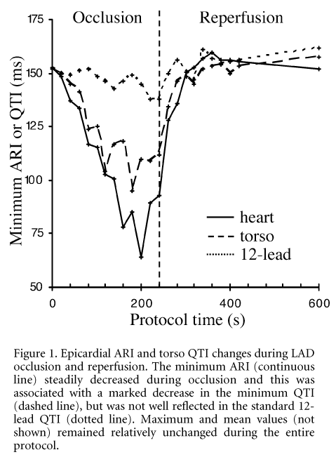
Diagnosing myocardial ischaemia with a standard 12-lead ECG may result in false negatives due to its poor spatial resolution. We correlated changes in torso and epicardial electrograms (EGs) during regional ventricular ischaemia in an anaesthetised (α-chloralose, 100 mg kg-1 I.V.) 29 kg pig that was artificially ventilated and thoracotomised. The animal was killed humanely at the end of the experiment. A suture snare was used to ligate the left anterior descending (LAD) coronary artery and an elasticized sock containing 127 unipolar electrodes was placed over the epicardium. The chest was reclosed and a vest containing 256 ECG electrodes was fitted to the torso. Simultaneous arrays of torso and epicardial EGs were recorded every 20 s during 4 min of LAD occlusion, followed by a period of reperfusion. Epicardial activation-recovery intervals (ARIs) and torso Q-T intervals (QTIs) were calculated from the EGs. Data were sampled at 2 kHz using a UnEmap data aquisition system and visualized using anatomically accurate computational models of the ventricular epicardium (Nash et al. 2001) and the porcine thorax (Nash et al. 2002).
LAD occlusion caused the minimum epicardial ARI to steadily decrease (see Fig. 1), whilst the location of this minimum shifted from the posterio-basal ventricular muscle (control) to the middle of the ischaemic region on the anterio-apical myocardium. These changes were associated with a steady decrease in the minimum torso QTI as it moved from the shoulder region (control) to the sternum. The 12-lead QTI was relatively unchanged. All electrical activity was fully restored following 6 min of reperfusion. We conclude that high spatio-temporal resolution torso mapping can detect cardiac ischaemia that is not always identifiable using indices derived from standard ECG limb leads.
This work was funded by The Wellcome Trust and the British Heart Foundation. The investigation was performed under a Home Office Project Licence PPL 30/1133.
All procedures accord with current UK legislation.

