
Physiology News Magazine
Obituary: Roger Yonchien Tsien MD, PhD
1952 – 2016
Membership
Obituary: Roger Yonchien Tsien MD, PhD
1952 – 2016
Membership
Timothy Rink, Cambridge, UK
https://doi.org/10.36866/pn.105.40
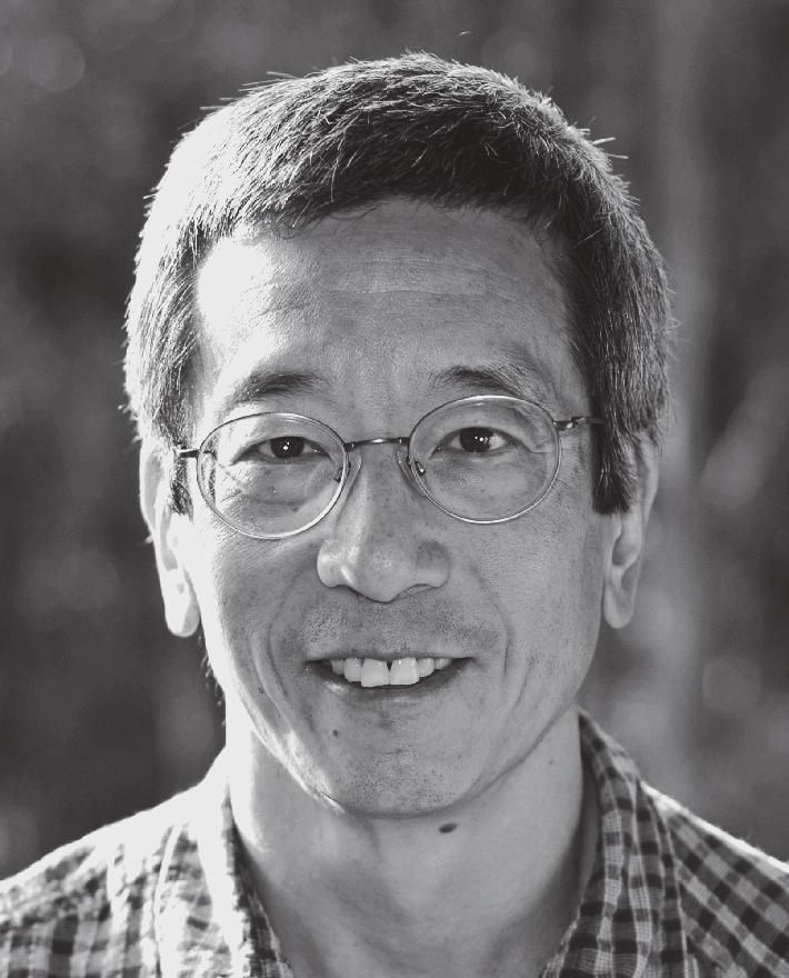
Roger Tsien died suddenly on 24 August 2016. He was a scientific and inventive genius who straddled traditional subject boundaries and shared the 2008 Nobel prize for chemistry for his work on the green fluorescent protein. Roger is best known for the extraordinarily diverse ‘optical toolkit’ of molecular probes that he created to study the physiology of intact living cells, many now used by thousands of scientists worldwide whose work they facilitated and transformed. His death at the age of 64 was a shock, not only to his family, but also to the scores of scientists who were trained by him and collaborated with him, and to the many who knew and used his work.
Childhood and early education
Roger Tsien was educated at Livingston High School, New Jersey and went up to Harvard aged 16, graduating at 20. He then moved to the Physiology Laboratory at the University of Cambridge for his PhD and postdoctoral research.
His parents were Chinese, his father an engineer who had done post-graduate studies at MIT before World War 2. He was later able to emigrate to the US in the aftermath of the war with his wife, a nurse, and their first son Richard. Roger developed a keen interest in chemistry from a very early age, and from the age of 8 performed increasingly complex, potentially hazardous, reactions in the home basement and garden with home-made glass apparatus and the then ubiquitous red rubber bungs. An amusing account of these activities is given in Roger’s ‘Nobel Biographical’. In 1967 he did an NSF sponsored summer internship at Ohio University on the ambident co-ordination of thiocyanate, which won him the $10,000 first prize in the prestigious nationwide Westinghouse Science Talent Search. In 1968 he entered Harvard and studied a wide range of subjects including art history, music, economics, relativistic quantum mechanics, astrophysics, molecular biology and neurobiology. Interestingly, he did not take chemistry because he considered those ‘ironically, the worst courses’. His first published paper was written in his final undergraduate year, in theoretical physics!
Doctoral research at Cambridge
Roger’s most abiding interest was neurobiology and the relationship between brain and mind. That led him to the Physiological Laboratory in Cambridge on a Marshall Scholarship. Strangely his assigned PhD advisor was Richard H Adrian, a distinguished muscle electrophysiologist who had no research expertise in neurobiology. But in those days PhD students were often left essentially to their own devices, with supervisors providing occasional ‘adult supervision’ and moral support. This allowed the student considerable freedom to operate in whatever area of research they chose. Roger did not even work in Adrian’s laboratory, but was allocated a windowless store room in the biophysics sub-department headed by Dennis Haydn. Early experiences with microelectrodes had convinced him to design and synthesise new dyes for recording nervous activity. To pursue his plan, he needed to learn synthetic organic chemistry. He persuaded two faculty members in Chemistry to guide him informally, and Roger commenced working at the one spare place in the Chemistry Department’s third year teaching laboratory. He related that once he was doing chemistry aimed at producing a probe he had designed to be useful, he enjoyed and became highly proficient in these syntheses.
His first attempt to make voltage sensing dyes by labelling tetrodotoxin was not successful. Roger then turned his attention to the design of dyes that could measure intracellular free Ca (Cai), and among other things report action potential activity indirectly. This would be a considerable challenge since Cai was known to be micromolar or less in the face of mM Mg, and much higher levels of Na and K. (By the early 70s others had managed to measure Cai with the luminescent protein aequorin or the organic dye Arsenazo III microinjected into large cells such as squid giant axons or barnacle giant muscle fibres.) Roger set out to design superior indicators based on the colourless Ca-selective chelator EGTA. This took quite some time to accomplish.
In the meantime he collaborated with me (then a young researcher recently come to Physiology and wanting to look at the role of Cai in stimulus-response coupling in cells where aequorin or Arsenazo would be unfeasible or inadequate) using his organic, inorganic and physical chemistry insights to design suitably sensitive and selective Ca-sensitive microelectrodes. As well as adapting a selective resin recipe from macroelectrodes, by for example adding PVC, we refined the production of the glass microelectrode itself. We cleansed micropipettes in ethanol-nitric acid, with some trepidation as this is an explosive mixture if not managed correctly, silanised them absolutely dry in an oven and painted them with silver paint to reduce capacitance, finally bevelling them on a rotating disc of diamond dust layered on an agar gel.
We had no adequate high impedance electrometers available to record from these ultra-high resistance microelectrodes, and so Roger promptly designed and built one to his own design (it was the topic of a communication to the Physiological Society) and it worked perfectly for at least a couple of decades thereafter. He saw nothing unusual in being able to accomplish this task; it was just another intellectual challenge to be met by some calculations, clear thinking, and maybe a focussed literature search. Working with various colleagues (including Richard W Tsien) we used these electrodes in skeletal and heart muscle, snail neurones and Xenopus embryos. This work was arduous, time resolution was poor, inapplicable to most mammalian cells and needed highly skilled researchers to perform the electrophysiology, which neither of us were.
So, further motivated, Roger pressed on with the work to design and synthesise fluorescent Cai reporters. His first successful fluorescent molecule was quin2 which was derived from a parent molecule, DAPTA, which gave only an absorbance, not a fluorescence signal. However the experience with Ca microelectrodes showed the critical need to design a method for introducing fluorescent probes, which were hydrophilic and membrane impermeant (as they needed to be, to stay in the cytosol as reporters) into populations of intact cells. Thinking through this problem and perusing the literature on making penicillins orally bioavailable, Roger made acetoxymethyl (AM) derivatives as a non-disruptive technique for loading hydrophilic molecules into cell. This technique for dye-loading intact cells soon became very widely used, and its invention was a major technical advance.
Having in hand quin2 and a method for trapping it in intact cells of any size, Roger and collaborators proceeded to make the first measurements of Cai in many cell types such as lymphocytes, platelets, sperm, neutrophils and macrophages. They started to explore the role, and sometimes the absence of a role, of Cai, in stimulus-response coupling, and also to investigate cellular regulation of Cai. This work led to numerous requests for samples of these probes, too numerous for him to supply with the available facilities.
To satisfy these requests Roger made two moves. First, he tried via the NRDC (the then mandated government agency that performed patenting and technology transfer for universities) to apply for patents, but they ruled that intracellular Ca was not commercially relevant (an error, though not so severe as later their failure to patent monoclonal antibodies) and they suggested he should work on probes for serum Ca. Second, he looked for a reagent company to synthesise and distribute the quin2 and quin2-AM to researchers world-wide. It turned out that these molecules were not that easy to produce and it took a while to find a company capable of doing a decent job. Lancaster Synthesis became the supplier and soon these probes became up to 50% of its business! Important further developments in Ca probes would come later, but while still at Cambridge, Roger designed and made a superior fluorescence cytosolic pH reporter, BCECF, which could be trapped in cells using the AM trick. In 1977 Roger finally wrote his thesis and was awarded a PhD.
From 1977 to 1981 he was a research fellow at Gonville and Caius College. He remained interested in fluorescence probes of membrane potential and worked with Steven Hladky and me on two classes of molecules that could report membrane potential due to redistribution across the plasma membrane. We worked out the physical chemistry of these systems in some detail and they gained moderately wide acceptance by cell biologists. However, they were not fast enough to report action potentials; Roger came back to that problem many years later.
Move to UC Berkeley
In spite of his accomplishments at Cambridge, and his manifest intellectual brilliance, Roger had difficulty in finding a permanent position. Cambridge had provided in many ways the ideal unstructured environment for him, with its permissive atmosphere and wide variety of visitors, to refine the many interdisciplinary skills he later exploited so well, but he was neither a ‘card-carrying’ biologist nor a ‘properly’ qualified chemist. In those days such boundary conditions were important to appointment committees; but after some searching, he received an offer to move as an assistant professor of Physiology and Anatomy to the University of California at Berkeley. Physiologists there such as Robert Zucker were keen to collaborate, and there were some, if primitive, facilities for doing his chemistry.
At Berkeley he was able to recruit synthetic chemists to his lab, one of whom, an ex-pat Englishman Stephen Adams, has worked with Roger throughout the remainder of his career. He continued to invent many further probes of intracellular ions, including a new generation of Cai probes (fura2, indo1, and fluo3) each of which had specific virtues. The most important was fura2 which was designed to give stronger signals than quin2, and also provided Ca-selective FRET (fluorescence resonance energy transfer)-based colour change. These improvements gave a probe that had much less background interference and much faster responses. Next came the development of intracellular Ca imaging for which Roger and his team were key pioneers. Among his many skills he was good at optics and computing and, working with Nikon, his team made one of the first systems for real-time, two wavelength, ratio imaging to research cell heterogeneity and Cai gradients within cells, and also rapid transients. His team also created the ratiometric Nai indicator SBFI.
Roger had a pithy phrase for research in cell signalling ‘show it, block it, move it’, using ‘spies, saboteurs and messengers’. With the aforementioned probes he had made major advances in ‘show it’. It had also been possible to trap enough DAPTA or quin2 into cells to greatly curtail Cai changes and thus to ‘block it’. Of course there were also pharmacological agents for blocking various signalling events. The Berkeley team produced a series of photo-labile Ca chelators, ‘caged calcium’, and collaborated with several neuroscience groups to study the role of Ca in control of neuronal ion channels, and with other groups in a variety of cell types. He also devised a photo-labile chelator that became more avid after illumination and so could locally reduce Cai. Roger’s practice at this time and henceforth was typically to publish a new probe by exemplifying its use to answer some significant biological question, either with expert collaborators, quite frequently local colleagues, or with some of his own post-grads and post-docs. As far as practical, Roger would then distribute probes to the scientific community from his lab or arrange that they were available from a commercial source.
Move to UC San Diego
Despite these continued successes, support facilities and funding at Berkeley were quite limited; so Roger decided to move to UCSD in 1989, where until his death he was professor of Pharmacology Biochemistry and Chemistry with a well-funded Howard Hughes Investigatorship. Inventions made early on at UCSD included intracellularly trappable forms of inositol tris and tetrakis phosphate – a far from trivial chemical challenge; a caged inositol tris phosphate, and a caged nitric oxide; and highly effective intracellularly trappable cAMP and cGMP. Given more facilities for chemistry and molecular biology he branched out into broader aspects of molecular and cell biology. In collaboration with Susan Taylor, Roger and his team devised an intracellular FRET probe for cAMP based labelling engineered versions of the regulatory and catalytic units of cAMP dependent kinase with different dyes
Roger had been seeking ways to use genetically encoded molecules to serve as his intracellular ‘spies’. He recognised that the majority of cell and molecular biologists were well versed in cloning, transfection and genetic engineering, rather than the somewhat arcane techniques of cell physiology, and that the broadest use of new tools would likely come from those that could be delivered to cells by suitable transfection methods. He was initially intrigued by the intensely fluorescent phycobiliproteins from cyanobacteria but the need for an accessory co-factor was a major drawback . He discovered in 1992 that the gene for the green fluorescent protein, GFP, from the jelly-fish that provided aequorin had just been cloned by Douglas Prasher. Roger greatly appreciated Prasher’s openness in supplying the GFP cDNA which allowed him and his colleagues to get to work rapidly. GFP was potentially highly attractive because it did not require an accessory co-factor. GFP produced by transfecting cells was intrinsically fluorescent. The development of many variants of GFP to probe multiple aspects of cell structure and function was a major turning point. Roger’s laboratory led in this venture as recognised by the 2008 Nobel prize, awarded to Roger together with Shimomora who first isolated GFP and Chalfie who pioneered its use in intact organisms to follow cell fate. Roger ensured that Prasher attended the Nobel celebrations and received much appreciation for his key contribution in first cloning the gene. The applications devised for GFP by Roger and his collaborators are far too numerous to even list here and in any case an excellent account of progress through 2008 can be found in the Nobel lecture. By a mixture of rational design and random mutagenesis many different coloured GFPs were generated (often called GFPs even if they glow actually blue or yellow). To further extend the ‘palette’ of colours Roger followed up the discovery of a new class of red fluorescent proteins by detailed investigation of their physical chemistry and the production of new longer wavelength colours and superior biophysical properties. He took great pleasure in naming these variant FPs after fruits of different colours, such as honeydew, tomato cherry and plum. One ingenious new tool he developed to produce superior variants of interesting proteins was to subvert the genetic apparatus in B lymphocytes that normally hypermutates antibodies in the process of affinity maturation.
A few of the applications of GFPs Roger’s team introduced to perform what he called ‘intracellular biochemistry are tools that allow study of: inositol tris phosphate, Cai, cAMP, cytosolic pH, specific protease detection, kinase and phosphatase activity, and mitosis. Each demanded thoughtful and careful choice of the engineered proteins to which the appropriate GFP would be attached. Another aim was looking at cell signals in specific sub-cellular compartments by targeting the probes to e.g. mitochondria, initially with his longstanding colleague Tullio Pozzan at Padua, who originally worked with Roger on quin2 in Cambridge days.
The astonishing success of the GFP family of sensors and the renown it brought did not inhibit or even slow down Roger’s desire to create yet more ways to ‘peek and poke’ into biology. One drawback of the GFPs is their large size that might interfere with the molecular events they are reporting. The solution was called FlasH, wherein a small peptide (much smaller the GFP) with four carefully positioned cysteines was the genetically encoded part of the probe that could be encoded in the DNA attached to that for any desired protein. The other component was a membrane permeant arsenical compound that could be applied to the transfected cells and becomes fluorescent only when complexed to the four SH groups.
In the 1990s he returned to devise better probes of membrane potential. With these probes he showed that one could follow fast action potentials with decent signal-to-noise, a step up from the previous two component system. He also worked out a way to make a FRET-based gene expression reporter that signalled from within intact living cells. This method provided a bacterial enzyme, beta-lactamase, which would have zero natural background in mammalian cells as the element to be inserted at the desired place in the genome of the cells to be studied. Then he and Gregor Zlokarnik invented a substrate with a FRET pair of fluorophores separated by a beta-lactam linker, as an acetoxymethyl ester so that it could be incorporated in living cells. Now expression of the gene in question would be reported non-destructively by ratiometric fluorescence in populations of cells or one cell at a time by imaging microscopy. This technology is now sold commercially as GeneBLAzer and has been used highly effectively in drug discovery for creating and screening cell-based assays.
With Stephen Adams, Roger developed a series of probes for protein localisation that gave both a light and electron microscope stain of the same molecules in the same sample to allow correlation at micrometre and nanometre scale. He also recently produced far red responsive variants of channel rhodopsins to extend the range of optogenetic technology for neurobiology.
The two projects that most engaged him in recent years were very different. One aimed at designing probes that targeted cancers in patients, as optical aids to surgery, MRI or positron emission imaging, or potentially as vehicles selectively targeting anti-tumour agents to the cancers. These were ingeniously designed molecular gadgets which on injection into a subject with cancer, were cleaved to produce either an increased signal due to de-quenching or a colour shift due to release of the FRET. Either way suitable imaging systems can show the operator (surgeon) where in the operating field is cancer and where the cancer-free margin is. After showing effectiveness in preclinical models, one such probe is now in clinical
trials with a San Diego biotechnology company, Avelas. Inc. Roger’s team also has preclinical data indicating that this approach can deliver anti-tumour drugs with increased therapeutic index.
The other big new project brought him back to the brain. He had long pondered what could be a basis for long term memory. He did not think that long term potentiation or synaptic and dendritic spine alterations could survive months and years as accurate long term engrams. As he read the neurobiology literature and pondered what long term store might be based on, he was intrigued by a structure with a set of proteins that are highly stable and closely connected with synapses and dendritic spines. This structure is the extracellular perineuronal net which surrounds neuronal processes, and indeed through holes in which dendritic spines must communicate. To summarise, he proposed the elegant, and in principle simple, concept that very long term memory stores are created by the number and pattern of these perforations (which for those old enough to recall he likened to a three-dimensional version of the punch cards we used in early computer programming up to about 1975). He adduced some interesting data that fitted with this idea and had embarked on a series of clever experiments to begin to support, or contradict, this radical notion. The requisite enzymes for making holes in the fibrillar network are present and subject to various biochemical controls. One neat point he raised is that the timing of the earliest of childhood memories fits roughly with the first development of the perineuronal net in infants. Even as a brilliant Nobel laureate he had difficulty getting any traction for this idea or support for the research. To publish the concept, he used his privilege as a member of the National Academy of Science to publish his hypothesis in PNAS. Unfortunately, now time, not Roger, will tell whether this is another example of aging Nobel laureates wanting to explain the brain, or a true breakthrough.
Roger was a brilliant research scientist. He is an author of over 400 papers and major reviews, with frequent publications in Nature and Science. He also did his fair share of reviewing papers and grants, supporting recruitment, and was also a fine educator and mentor, and trained over 100 graduate students and post-docs from many different disciplines. While he was an introverted, somewhat shy person, he was a superb lecturer using everyday plain language to explain complex ideas and avoiding jargon. He also had wonderful and colourful slides and videos and would many times give colourful practical demonstrations like the famous chemistry lecturers of old. He was a wry and insightful participant in Q&A after presentations or in symposia. His breadth of knowledge and expertise meant there were few presentations the he could not cannily interrogate no matter what the field of biology and indeed beyond. He did not wish to be distracted from the science that he and his group were doing and so did not take on administrative tasks such as a departmental or faculty chair. His achievements were recognised by dozens of prizes and awards and scores of plenary or keynote lectures. Aside from the Nobel Prize, other major awards included: Javits Neuroscience Investigator; Gairdner Foundation International; Foreign Membership of the Royal Society; HP Heineken Prize; Max Delbruck Medal; Wolf Prize; Golden Goose Award.
In addition to his tireless pursuit of academic excellence, Roger was a master translational scientist, before that became usual (or even acceptable) and encouraged. There are over 160 US patents in which he is named (and usually was the lead) inventor. While he was naturally keen to participate in or lead the first application of his new tools and produce the first publication he was generous in providing materials to the scientific community. Many of his reagents were manufactured and distributed by licensees of the patents, in particular Molecular Probes of Eugene OR, which was founded and run for years by a creative chemist, Richard Haughland who became a valued colleague, if occasionally also a competitor, of Roger. Roger consulted for many biotechnology and pharmaceutical R&D groups over the years and was founder or co-founder of three San Diego companies. He once semi-seriously commented that one motivation for founding these companies was to provide good jobs for his post-docs. He recognised that the kind of innovative cross-disciplinary science he got them doing in his labs was great training for the challenges of high level applied research in science-based industrial ventures. One company’s biggest success, coming to fruition after it became part of Vertex Inc., where Roger remained on the scientific advisory board, was the launch of two drugs that enhance the defective chloride channel in cystic fibrosis. With his colleague Charles Zuker and the renowned biochemist Lubert Stryer, he also co-founded Senomyx in 1998, which aimed to use the new knowledge of taste receptors and the developments of the Tsien technologies applicable to compound screening to discover food additive compounds that could potently modulate taste receptors to for example, enhance sweet flavour or block bitterness. More recently he founded Avelas Biosciences to develop the cancer illuminating structures mentioned above.
Roger worked hard and intensively at his science. At home in the evening he would often sit cross-legged on the floor after dinner, with a weather eye perhaps on some TV drama, but his ever-present note book on his lap, pencil in hand, designing the next probe, or deep in a biophysical calculation. Away from work, Roger applied himself quite intensively to his other interests. He was a fine pianist and had briefly even considered a musical career. As soon as he had the space and funds, he acquired his own grand piano. He travelled with a Walkman, or modern equivalent loaded with the classics. He enjoyed somewhat down-market thrillers – John D MacDonald was a favourite author. Fitting with his joy in colours and imaging, he was a gifted amateur photographer, and enjoyed vacations in the wild outdoors, taking arduous treks, camera in hand.
In 2014 he suffered a stroke that left him with significant disabilities including a right hemi-paresis and some minor dysphasia. His intellect was not impaired, and with determined efforts in rehabilitation he was able to continue his science and inventions and indeed to travel and continue to give outstanding keynote lectures. But it was burdensome and tiring, and he had begun to wind down his research team in last year or so. He and his wife Wendy moved from San Diego to a delightful spot outside Eugene to start a new quieter existence. Sadly, one year after the move, Roger died while exploring on his tricycle in a local park.
Roger was for many the brightest person they ever met. He was generous with his time and intellect and in his own way totally non-elitist, not that he suffered fools gladly, especially if they had position and pretensions. But he was very supportive of motivated young scientists and had a major impact on so many careers, naturally those who worked for or with him, but also many thousands whose work was enabled by his inventions. He was very special, touched the work and lives of many, and will be missed by those who knew him, as a generous friend, and for his wonderful mind. Roger is survived by his wife Wendy, and his two brothers Richard and Louis.