The vascular contributions to cognitive impairment and dementia (VCID) have been recognised as a research priority [1]. Current treatments for cognitive deficits in Alzheimer’s Disease centre on rescuing cholinergic transmission with acetylcholinesterase inhibitors, for example donepezil. Importantly, there is evidence that donepezil can also have a benefit on cognition in vascular dementia (VaD) and patients with vascular cognitive impairment [2-4], indicating that the cholinergic system can directly impact on cognition in cerebrovascular disorders. Of the five muscarinic acetylcholine (ACh) receptor subtypes (M1-M5), the M5 receptor has been suggested to play a role in cerebral blood flow (CBF) regulation [5,6]. Here, it was hypothesised that M5 receptor expression would be enriched in the cerebrovasculature of rat brains.
Whole brain (WB) homogenates from stroke prone spontaneously hypertensive rats (SHRSP), underwent a vessel enrichment protocol using a ficoll gradient. Cerebral and peripheral vessels were also dissected from SHRSP, namely the middle cerebral artery, basilar, transcending aorta, carotid and mesenteric arteries. RNA was isolated from WB homogenates, vessel enriched fractions (S1) and individual vessels. qPCR used to determine the expression levels of M5 & M3 muscarinic receptors along with claudin-5 tight junction protein and aquaporin 4 (AQP4) (expressed predominantly on astrocyte end-feet) relative to the housekeeping gene b-actin. Data was analysed using a paired t-test and p<0.05 was considered statistically significant.
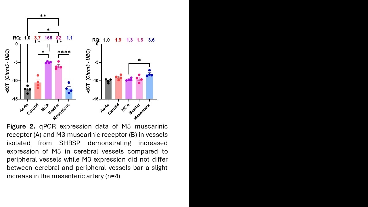
M5 expression was significantly 66% higher in S1 fractions compared to WB (p=0.01) while M3 expression appeared to increase by 95% although this was not statistically significant (p=0.08) (Figure 1). Claudin-5 and AQP4 were significantly increased in S1 fractions by 1800% (p=0.0007) and 65% (p=0.03) respectively, confirming vasculature enrichment in the samples. Further to this, M5 expression was significantly between 82 and 166-fold (p<0.01), 22 and 45-fold (p<0.05), and 74 and 150-fold (p<0.01 and p<0.0001) greater in the MCA and basilar artery of the SHRSP rat compared to the aorta, carotid and mesenteric arteries, respectively (Figure 2). M3 expression, however, was similar across all arteries assessed with the exception of a 3-fold increase in expression in the mesenteric vs the basilar artery (p<0.05).
In SHRSP brains, the M5 muscarinic receptor expression is significantly enriched in the cerebrovasculature in both vessel enriched brain fractions and when assessing expression in dissected vessels. This may suggest an important role of M5 in the cerebrovasculature which may provide a potential novel therapeutic target for improving CBF in cerebrovascular disorders and VaD.