Background
Neuroinflammation plays an important role in the development and progression of neurodegenerative disorders, particularly Alzheimer’s disease (AD) and Parkinson’s disease (PD). Genome-wide association studies indicate that many inflammatory genes associated with AD are exclusively expressed in microglia. AD is the leading cause of dementia and is characterized by the accumulation of extracellular amyloid-β plaques and intracellular neurofibrillary tau tangles. However, clinical evidence suggests that cases of ‘pure’ pathology involving only amyloid-β and tau are relatively rare. More than 50% of AD patients also show accumulation of TDP-43, while approximately 33% exhibit accumulation of α-synuclein. This highlights the importance of concomitant pathology in AD.
Aims
The project aims to investigate how concomitant pathology affects microglia. It focuses on the development of methods to enrich pathogenic aggregated proteins from patients’ brain samples, treating Induced pluripotent stem cell (iPSC) – derived microglia with the aggregates, and investigating the mechanisms behind neuroinflammation leading to neurodegeneration in AD.
Methods
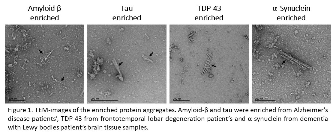
We have standardized the isolation of amyloid-β, tau, TDP-43, and α-synuclein from brain tissue of patients in the Oxford Project to Investigate Memory and Ageing (OPTIMA) cohort. We have characterized the isolated aggregates using Western blotting, transmission electron microscopy (TEM), and seeding bioassays. Isolated aggregates were quantified using ELISA and mass-spectrometry. iPSC-derived microglia are being treated with combinations of the isolated aggregates to explore how this affects the genetic and proteomic landscape of these cells. Human biological samples were sourced ethically, and their research use was in accord with the terms of the informed consents under an IRB/REC (number 23/SC/024) approved protocol.
Results
Western blot and TEM (Figure 1) demonstrate isolation of pathogenic aggregates. Using a seeding bioassay, we have been able to confirm that isolated aggregates are seed competent. Comparison of several suppliers’ ELISA kits shows them to give widely divergent results for the same samples.
Conclusions
We have standardized the isolation of seed competent aggregates from small-scale tissue samples. Treating microglia with different combinations of these aggregates will enhance our understanding of how mixed pathology influences the development and progression of AD and may help identify new targets for the potential treatment and management of the disease.