The external appearance of uniformity of cardiomyocyte contractions emerges from coordination of numerous, spatio-temporally heterogeneous processes, involving specialised sub-cellular nano-domains. Through omnidirectional coupling of electrical activity, calcium fluxes, and mechanical action, cardiomyocytes rapidly adjust to ever-changing physiological demand. It remains unknown how dynamic ultrastructural cross-talk contributes to the marked cardiac auto-regulatory capacity.
I propose that beat-by-beat auto-regulation of heart function is governed at the nano-scale by a process unique to cardiomyocytes: the rhythmic cycle of cardiomyocyte stretch, contraction, and relaxation. In this concept, the heartbeat is not merely the consequence of cardiomyocyte activity, but an intrinsic regulator that continually modifies nano-domain structure and function in preparation for the next beat.
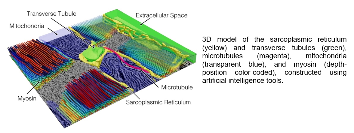
In order to understand beat-by-beat cardiomyocyte nano-dynamics, we combine time-resolved 3D electron tomography, sub-cellular functional imaging, and artificial intelligence-assisted segmentation of cardiomyocyte sub-cellular ultrastructure, where deep learning-based segmentation enables automated reconstruction of complex cardiomyocyte ultrastructural components. Finally, we use artificial intelligence-based generative models and computational modelling for data synthesis, and for testing and generation of new hypotheses.
We have shown that cardiomyocyte contraction and stretch are associated with beat-by-beat deformation of cardiomyocyte organelles: transverse-axial tubular system (TATS), sarcoplasmic reticulum, mitochondria, microtubules, caveolae, and nuclei. In order to visualise cardiomyocyte contraction on the nano-scale (something that would be impossible experimentally) we used generative adversarial models to synthesise biophysically constrained ‘motion pictures’ of cardiomyocyte ultrastructure with nanometre-resolution, creating a powerful hypothesis-generation platform linking nanoscale structural deformation to electro-mechanical function.
As an example of a functional relevance of organelle deformation, we have shown that TATS (a network of surface membrane invaginations that enables close structural coupling of plasma membrane and extracellular fluid with sarcoplasmic reticulum) cyclically deforms during cardiomyocyte stretch and contraction in rabbit (all investigations conformed to national and international animal welfare laws). Cyclic, beat-by-beat TATS deformation which is associated with faster luminal content exchange with the bulk extracellular space, an effect that scales up with contraction amplitude and frequency. TATS ‘pumping’ serves to abolish intraluminal calcium depletions that would otherwise develop as a consequence of transmembrane calcium fluxes, and – if left unrectified by deformation-driven pumping – would lead to the loss of the contractile force.
In conclusion, our studies represent an innovative framework that can provide unique insights into the nanoscopic dynamics of cardiomyocytes. Cardiac organelle mechano-sensitivity likely represents a fundamental principle of cardiac physiology, where mechanical modulation of nano-domain structure and function can help us understand how cardiomyocytes adapt rapidly to changes in circulatory demand.