
By Tashi Raina and Alireza Mani from University College London (UCL), UK
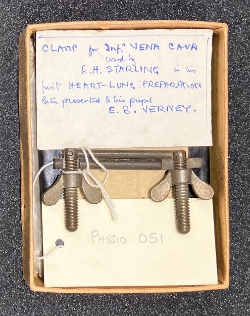
In the UCL Historic Objects and Collections series, the team explores the stories behind the physiological artefacts housed within the Institute’s rich collection. The object featured in this blog is Physiological Instrument #051, a seemingly ordinary clamp, housed in the UCL Object-Based Learning Laboratory. This clamp was used by Ernest Starling in his heart–lung preparation, developed at UCL in the early 1910s. Starling later presented the clamp to his pupil, Ernest Verney, who developed the heart–lung–kidney preparation and went on to discover the hormonal regulation of blood osmolarity through the action of antidiuretic hormone.
The clamp
At first glance this clamp is quite unremarkable, sitting amongst the other artefacts within UCL’s science collection. Even when placed alongside any number of other clamps from its time period, it would be quite indistinguishable. However, this particular clamp has a significant chapter of physiological history bound to it (Figure 1).
Evolution of heart-lung preparation
During the late 1800s scientists aimed to deepen understanding of the individual function of organs and how they worked within the physiological system. Carl Ludwig (1816-1895) is considered to be one of the fathers of experimental physiology and the pioneer of isolated organ perfusion; where an organ can function outside of its organism’s body or segregated from its default physiological connections. In 1846, Ludwig and his students at the Leipzig Institute of Physiology (Germany) developed the earliest isolated heart through connection of the aorta of the excised organ, to the carotid artery of a living donor animal, and maintained the isolated heart’s pumping capacity (1). H. Newell Martin (Johns Hopkins University, US) established the first mammalian heart-lung preparation in 1883 (1), and this was further improved by Oscar Langendorff in 1895 through developments in retrograde perfusion of the isolated heart (2).
Preventing blood clotting
The heart-lung preparation involved a deeply anaesthetised animal that was hooked up to a ventilator to help it breathe. The circulation between heart and lung remained untouched however the rest of the systemic circulation was severed. Other major blood vessels were temporarily clamped, incised and a cannula added to redirect the blood into a reservoir. Perfusion of isolated organs required blood; however, once blood left the intact vascular system, it rapidly clotted through the formation of fibrin. Heparin had not yet been discovered (it was first identified in 1916), and the only known anticoagulant, hirudin, extracted from medicinal leeches, was not readily available.
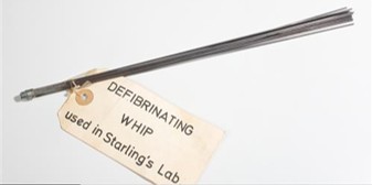
Physiologists therefore developed an alternative method to prevent clotting by mechanically removing fibrin from freshly collected blood, producing what became known as defibrinated blood. The blood to be circulated was diverted out of the animal, whisked using a tool known as a defibrinating whip (Figure 2) and then redirected back into the experimental system’s circulation (3). This prevented fibrin from obstructing vasculature and tubing which enabled smooth passage. The heart could now be studied under experimental conditions.
Starling’s laboratory
Ernest Starling (1866-1927) was one of the world’s leading physiologists, renowned for the groundbreaking research he conducted at UCL. Like Carl Ludwig’s celebrated Leipzig Institute, his laboratory attracted scientists from around the world and provided an environment in which young investigators could learn, collaborate, and make their own discoveries. Starling was particularly interested in how organs communicated to regulate the body as a whole. Working together with William Bayliss in 1902, they discovered that the intestine could stimulate the pancreas by the release of a chemical messenger, secretin, into the bloodstream (4,5). This challenged the prevailing Pavlovian theory of the time, where the nervous system was the ultimate regulator of biological and behavioural homeostasis (6). The line of questioning then further shifted to the other organs that were potentially affected by hormonal actions, one being the kidney.
The kidney
By 1900, physiologists had deftly begun exploring the kidney. They understood the structures of nephrons and glomeruli, renal circulation and the filtration processes that lead to urine formation. Whilst the kidney was understood anatomically and in a mechanical sense it was unclear how the kidney was controlled within the functions of the entire organism. Decisions regarding urine concentration, dilution and sensing dehydration were yet to be attached to a physiological concept of either independent kidney function, nervous system regulation or part of a hormonal system. The next breakthrough would come from a relatively unknown, yet exceptional, pupil of Starling, Ernest Verney (1894-1967).
Ernest Verney and the heart-lung-kidney preparation
Verney joined Starling’s laboratory at UCL in 1921, where he gained access to a culture of rigorous experimentation as well as guidance under Starling himself. Prior to Verney’s arrival, Starling inherited the earlier heart-lung techniques developed by Newell Martin and Ludwig and was proposing adaptations to involve the kidney. In 1914, just before the First World War, Starling attempted the first heart-lung-kidney preparation; however, it was mostly a failure due to lack of urine production from the kidney. Physiologists were then diverted towards war-time work, which halted the progress on the kidney project (6). Once the war had ended, Starling resumed the project in 1921, working alongside Verney.
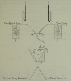
Starling and Verney built upon the heart-lung-kidney model and this time preserved the arterial supply to the kidneys through the abdominal aorta and renal arteries to maintain perfusion (Figure 3). Any vasculature supplying other organs was clamped, the blood within redirected to an external reservoir, defibrinated and then returned to the heart-lung-kidney circuit. The addition of the kidney to the original model was incredibly complex; with more clamps, cannulas and blood required to ensure efficacy (7,8). They injected posterior pituitary extract (pituitrin) into circulation and observed urine volume falling and concentration increasing (9).
The new model allowed in depth study of urine production filtration, tubular secretion and reabsorption as well as evidence that the presence of a hormone circulating in the bloodstream controlled the kidneys’ water excretion. They published their findings in a 1922 paper in The Journal of Physiology, 20 years on from the first hormone being discovered (7). Following the first successful use of the heart–lung–kidney preparation, Starling and Verney published a series of influential papers (8,9) which laid the foundations for Verney’s later discovery of the hormonal regulation of water balance by antidiuretic hormone (10).
Starling and Verney had deduced the presence and effect of a hormone; however, its identity was still unknown. Tragically, Starling passed away in 1927 aboard a steamship in Jamaica; on a trip intended to restore his declining health. He left behind a legacy of numerous physiological discoveries: the Starling equation, the law of the heart and hormones. In addition to his discoveries, Starling presented a clamp to his pupil Verney, by way of a gift (Figure 1). The clamp served as a silent assurance from mentor to student, a sign of capability and respect, and is now archived at the UCL Objects-Based Library (Object LDUSC-PHYSIO-52).
Antidiuretic hormone
Verney spent the next 25 years investigating and attempting to demonstrate that the hypothesis of a hormone controlling kidney function was indeed true. In this time significant changes were made to the original heart-lung-kidney preparation. Verney recognised how anaesthesia affected blood pressure and urine production when given to the test subject animals. He adapted this by implanting cannulas while the animals were anesthetised and performed the actual experiment once the animal had woken up and recovered. This removed a source of experimental error. Verney also determined that change in osmolarity only provoked a change in kidney function if the change was detected by the brain. This indicated that the kidney was not deciding but responding (10).
Verney devised an experiment to demonstrate brain involvement through infusing hypotonic saline solution to the arteries supplying the brain, whilst keeping the kidneys exposed to relatively normal levels of blood. This led to the kidneys retaining water, despite never contacting abnormally concentrated blood (10). After deducing the brain’s control over water balance, alongside the understanding of posterior pituitary extract causing antidiuretic kidney effects, it became apparent that the pituitary gland directly impacted kidney function. Subsequent work by Verney and other physiologists determined that the pituitary is a storage and release site, but not where the blood concentration is monitored or where the hormone is produced (11). Verney proposed the existence of specialised sensory cells within the hypothalamus that responded to changes in blood solute concentration, pressure and volume. The change was relayed to the hypothalamus which then modulate the posterior pituitary, releasing the hormone into the blood (10).
Chemical structure of antidiuretic hormone
Verney and Starling’s work revolved around physiological function and branched into the theory of hormones, however the actual chemical identity of the antidiuretic hormone remained a mystery. This was where Verney’s expertise ended; a shift from physiological discovery to biochemical, led by Vincent du Vigneaud (1901-1978) and his research team at Cornell University Medical College, US. Over the time period of these extensive physiological discoveries, biochemistry had been catching up. Protein purification techniques, chromatography and amino acid analysis all became more refined and accurate.
Du Vigneaud himself was a specialist in peptide chemistry, and his laboratory focused on determining the chemical structure of the mystery hormone. This endeavour took enormous effort; thousands of animal pituitary glands were extracted, each with a minuscule quantity of hormone, just to collect enough to purify and study. Du Vigneaud then chemically broke the peptide into fragments and identified the amino acids. This mystery hormone, theorised about in 1925 and finally chemically identified in 1953, was made up of nine amino acids, with a characteristic disulfide bond linking two cysteine molecules. Du Vigneaud went on to synthesise antidiuretic hormone as well as oxytocin in his laboratory and was awarded the Nobel Prize in Chemistry in 1955 (13).
Although only a simple clamp, the instrument that was given to Verney symbolises far more than a piece of laboratory equipment. It reflects the collaboration, mentorship and continuity of scientific discovery, where each generation builds on the work of those before it.
Acknowledgment: The authors are grateful to Alanna Orpen (The Physiological Society), Ignacio Echeverria Faccin, Graham Isted and Liz Blanks (UCL Museums & Cultural Collections) for their collaboration and expert advice.
If you missed the first six blogs by the UCL Historic Objects and Collections team, read their blogs, Bárány’s Box, the Kymograph, Haldane apparatus, From Wills’ factor to folic acid, From squid giant axons to recording action potentials inside a nerve fibre, Measuring the invisible: The Hartridge Reversion Spectroscope and Winifred Cullis: Bringing human physiology to the public to discover more about the history of physiology.
References
- Zimmer H-G. (1998) The Isolated Perfused Heart and Its Pioneers. Physiology 13 (4): 203–10. https://doi.org/10.1152/physiologyonline.1998.13.4.203.
- Langendorff O. (1895) Untersuchungen Am Überlebenden Säugethierherzen. Pflüger, Archiv Für Die Gesammte Physiologie Des Menschen Und Der Thiere 61 (6): 291–332. https://doi.org/10.1007/bf01812150.
- Editors (1935) Maintenance of Life in Isolated Animal Organs. Nature 135:1066–1067.
- Bayliss W.M., and Starling E.H. 1902. The Mechanism of Pancreatic Secretion. The Journal of Physiology 28 (5): 325–53.
- Starling E.H. (1905). The Croonian Lectures on the Chemical Correlation of the Functions of the Body. The Lancet, 166, 339–341; 423–425; 501–503; 579–583.
- Henderson J. (2005) Ernest Starling and the Poetics of Physiology. Oxford: Oxford University Press.
- Verney E.B. & Starling E.H. (1922) On secretion by the isolated kidney. Journal of Physiology, 56(5), 353–358.
- Starling E.H., and Verney E.B. (1924) Die Folgen Der Trennung von Glomerulus- Und Harnkanälchentätigkeit Bei Der Säugetierniere. Pflügers Archiv Für Die Gesamte Physiologie Des Menschen Und Der Tiere 205 (1): 47–50.
- Starling E.H. and Verney E.B. (1925) The secretion of urine, as studied on the isolated kidney. Proceedings of the Royal Society B, 97, 321–363.
- Verney E.B. (1947) Croonian Lecture – The Antidiuretic Hormone and the Factors Which Determine Its Release. Proceedings of the Royal Society B, 135 (878): 25–106.
- Harris G.W. (1955) Neural Control of the Pituitary Gland. London: Edward Arnold.
- du Vigneaud, V., Lawler H.C., and Popenoe E.A. (1953). Enzymatic cleavage of glycinamide from vasopressin and a proposed structure for this pressor-antidiuretic hormone of the posterior pituitary. Journal of the American Chemical Society, 75(19), 4880–4881.
- du Vigneaud, V. (1955) Nobel Lecture: ‘Trail of Sulfur Research: From Insulin to Oxytocin. https://www.nobelprize.org/uploads/2018/06/vigneaud-lecture.pdf