

From squid giant axons to recording action potentials inside a nerve fibre
By Tashi Raina, Hannah Cornish and Alireza Mani from University College London (UCL), UK
The Historic Objects and Collections at UCL series presents a model made of wax representing the ganglion of a squid from the collection of the UCL Grant Museum currently on display at the UCL Object-Based Learning Laboratory, London. The object featured in this blog is a model created from the work of John Zachary Young, a UCL Professor of Anatomy from 1945 till 1974. It showcases his research on squid giant axons, which inspired Alan Hodgkin and Andrew Huxley to record the first action potentials from inside a nerve fibre.
Uncovering the nature of nerve signals
With a donut shaped brain, three hearts and blue blood; the squid is a fascinating cephalopod – but did you know of its significant contribution towards science? In the early twentieth century, scientists faced a fundamental mystery; how did nerves transmit signals? The human nervous system fires hundreds of signals every second however for much of modern scientific history, the precise mechanism of transmission remained entirely unknown. The answer eventually arrived not from a human specimen or a laboratory animal – but instead from the dark waters of the English Channel, within the mantle of a common squid.
As early as the 1780s, the Italian physician Luigi Galvani had demonstrated that nerves and muscles were electrical in nature. His now-famous experiments in which the legs of a dissected frog twitched when touched by a charged metal probe, established that animal tissue was stimulated by electricity. This idea earned Galvani both ridicule and reverence in equal measure. By the mid-nineteenth century, the German physiologist Emil du Bois-Reymond had cemented this insight, demonstrating that nerves generated measurable electrical signals; what he termed the negative variation – today recognised as the action potential (1). Despite these incredible discoveries, a mechanism had still not been established.
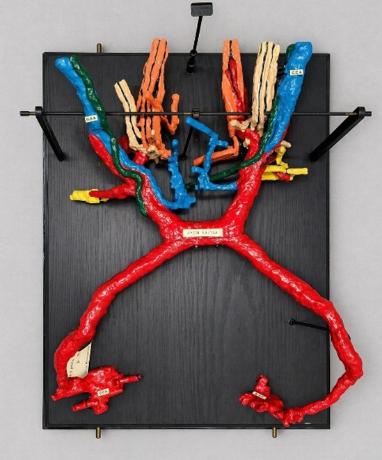
Figure 1. Model of giant fibres in the central nervous system of a squid, seen from above. Made from painted wax, with metal struts and a black wooden base (UCL Grant Museum of Zoology object LDUCZ-R250 on display at the UCL Object-Based Learning Laboratory).
Nerve fibres are the axons that carry electrical signals between cell bodies. They are extraordinarily small and fine structures, only a few micrometres in diameter, making measuring electrical changes inside the cell membrane impossible using rudimentary twentieth century equipment. Electrodes were too large and crude to be inserted within the nerve fibre without destroying it, forcing researchers of the time to record electrical activity from outside of nerves. This produced signals that were blurred by surrounding tissue and had weak resolution. The nerve’s interior – where the electrical events actually occurred – remained inaccessible.
In 1936, British zoologist John Zachary (JZ) Young was studying the nervous systems of squid at the Marine Biological Association. His field of interest was evolutionary, questioning how nervous systems had diversified across species. During dissection of squid caught in the Stazione Zoologica in Naples (Italy), Marine Biological Laboratory in Woods Hole (USA) and later in Laboratory of the Marine Biological Association in Plymouth (UK), he made observations that redirected the course of neurophysiology (2).
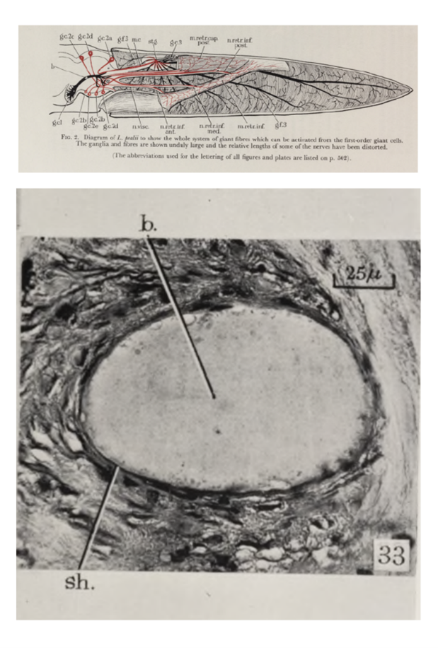
Figure 2. Upper panel: A schematic diagrams of giant fibres in Loligo squid from his 1939 publication in Philosophical Transactions of the Royal Society of London (3). The original illustration of the upper panel is on display at the UCL Grant Museum of Zoology. Lower panel: A section through the axonal bridge of Loligo squid in JZ Young’s 1939 paper (3). He noted only a single axoplasm (b) and a single sheath (sh.), indicating that the structure represents a true single axon rather than a combination of separate nerve fibres.
Discovery of giant axon in common squid
Running through the nerve bundle of the squid’s mantle – a muscle complex that powers the squid’s jet propulsion – Young discovered a singular, pale enormous nerve fibre, visible to the naked eye (Figure 2). Although it was roughly the thickness of the lead from a mechanical pencil; it was gargantuan compared to the microscopic fibres from humans. A typical mammalian axon measures <20 micrometres across, compared to the half a millimetre diameter of the squid nerve fibre. The fibre was termed the squid giant axon; and was part of the squid’s jet propulsion system, triggering the contraction of mantle muscles to expel water for quick escapes from predators. The nerve fibre achieves this because its internal resistance of the wider axon is lower, allowing impulses to travel at a greater speed than in a narrow fibre. In a 1939 paper Young described the giant fibre and argued that it allowed for a unique experimental opportunity (3).
Recording action potential from the giant axon
The invitation was taken up by two physiologists at Cambridge: Alan Lloyd Hodgkin and Andrew Fielding Huxley. In the summer of 1939, they travelled to the Marine Biological Association in Plymouth to work with the squid giant axon directly, alongside Young’s collaboration. Hodgkin and Huxley threaded a fine glass electrode longitudinally along the interior of the giant fibre and for the first time, a living nerve membrane could be measured from within. The results were immediate and unusual. When the nerve fired, the electrical potential across the membrane did not drop from its resting value toward zero, as the prevailing theory had predicted. Instead, it dramatically overshot, swinging from a resting potential of approximately -50mV to a peak of around +40mV before returning (Figure 3). The overshoot was significant, and implied that nerve transmission was not a case of passive conduction and instead was an active process where ions moved across the membrane in a sequence. A simple wire conducting electricity would not behave in this manner. Hodgkin and Huxley published their initial findings in Nature in 1939 (4). Then progress was interrupted by the war.
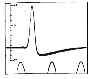
Figure 3. Action potential recorded between inside and outside of axon in Hodgkin and Huxley experiment published in Nature 1939 (4).
The Second World War
The Second World War saw the Plymouth experiments suspended immediately. JZ Young set up a research unit at the University of Oxford to study how damaged nerves regenerate, motivated by the large number of nerve injuries in soldiers (5). Hodgkin worked in the British Air Ministry towards developing airborne radar systems, and Huxley contributed to research on anti-aircraft gunnery. Hodgkin and Huxley spent the war years immersed in engineering problems and precision electronics and interestingly, the wartime work gave both a greater familiarity with analogue electrics and mechanical systems which they applied once back in Plymouth after the war.
Hodgkin and Huxley faced a technical problem within their investigation. When a nerve fires, the membrane voltage and the ionic currents change simultaneously and to understand what the currents were actually doing, they needed to freeze one variable while measuring the other. The solution was the voltage clamp, a feedback-controlled electronic circuit that held the membrane potential at a fixed, chosen value while the resulting ionic currents were measured directly. The voltage clamp worked by detecting any deviation from the set membrane voltage and instantly injecting a compensating current to cancel it out. This injected current was equal and opposite to the ionic current flowing across the membrane – and therefore a direct measurement of it.
The Hodgkin and Huxley model
Between 1949 and 1952, using the voltage clamp on squid giant axons at Plymouth, they published a landmark series of papers in The Journal of Physiology (6-12). These papers unpicked the ionic basis of the action potential. They provided experimental evidence that the rising phase of the action potential (depolarisation) was driven by the rapid opening of sodium channels, allowing sodium ions to flood into the cell along their electrochemical gradient. The falling phase (repolarisation) resulted from the subsequent opening of potassium channels, allowing potassium ions to exit the cell and restore the resting potential.
The culmination of this concept was the Hodgkin-Huxley model, published in their final 1952 paper (12). They encoded their findings into a set of differential equations that mathematically described the behaviour of the membrane conductance over time. These equations could be used to predict the shape of the action potential with a certainty that matched experimental recordings with remarkable accuracy. At a time when computational simulation of biological systems was almost unheard of, the model represented a quantitative and mechanistic description of a physiological process. The Hodgkin-Huxley model remains foundational even today.
In 1963, Alan Hodgkin and Andrew Huxley were awarded the Nobel Prize in Physiology or Medicine, shared with the Australian neurophysiologist John Carew Eccles, for their discoveries concerning “the ionic mechanisms involved in excitation and inhibition in the peripheral and central portions of the nerve cell membrane”.
The squid giant axon was one of the most productive experimental preparations in the history of physiology. What began as a scientific curiosity became the foundation of modern neuroscience. JZ Young was looking at squid and discovered a model system for electrophysiology. The giant axon was a consequence of evolution, optimised for predator evasion; and happened to solve a technical problem that had stalled neurophysiology for decades.
Acknowledgment: The authors are grateful to Emilia Kingham and Ignacio Echeverria Faccin (UCL Museums & Cultural Collections) for their collaboration and expert advice.
If you missed the first four artefacts on display by the UCL Historic Objects and Collections team, read their blogs, Bárány’s Box, the Kymograph, Haldane apparatus and From Wills’ factor to folic acid to discover more about the history of physiology.
References
1.du Bois-Reymond, E. 1848. Untersuchungen über thierische Elektricität. Berlin: G. Reimer.
2.Jones, K.M., 2022. ‘You’ve got to work on this axon’: J.Z. Young and squid giant axon preparations in 20th-century neurobiology. Berichte zur Wissenschaftsgeschichte, 45(3), pp.317–331. Available at: https://doi.org/10.1002/bewi.202200021
3.Young, J.Z., 1939. Fused neurons and synaptic contacts in the giant nerve fibres of cephalopods. Philosophical Transactions of the Royal Society of London. Series B, 229, pp.465–503.
4.Hodgkin, A.L. and Huxley, A.F., 1939. Action potentials recorded from inside a nerve fibre. Nature, 144, pp.710–711.
5.Young, J.Z. and Medawar, P. (1940) ‘Fibrin suture of peripheral nerves’, The Lancet, 236, pp. 126–128.
6.Hodgkin, A.L. and Huxley, A.F., 1945. Resting and action potentials in single nerve fibres. The Journal of Physiology, 104(2), pp.176–195. Available at: https://doi.org/10.1113/jphysiol.1945.sp004114
7.Hodgkin, A.L. and Huxley, A.F., 1947. Potassium leakage from an active nerve fibre. The Journal of Physiology, 106(3), pp.341–367. Available at: https://doi.org/10.1113/jphysiol.1947.sp004216
8.Hodgkin, A.L., Huxley, A.F. and Katz, B., 1952. Measurement of current–voltage relations in the membrane of the giant axon of Loligo. The Journal of Physiology, 116(4), pp.424–448. Available at: https://doi.org/10.1113/jphysiol.1952.sp004716
9.Hodgkin, A.L. and Huxley, A.F., 1952a. Currents carried by sodium and potassium ions through the membrane of the giant axon of Loligo. The Journal of Physiology, 116(4), pp.449–472. Available at: https://doi.org/10.1113/jphysiol.1952.sp004717
10.Hodgkin, A.L. and Huxley, A.F., 1952b. The components of membrane conductance in the giant axon of Loligo. The Journal of Physiology, 116(4), pp.473–496. Available at: https://doi.org/10.1113/jphysiol.1952.sp004718
11.Hodgkin, A.L. and Huxley, A.F., 1952c. The dual effect of membrane potential on sodium conductance in the giant axon of Loligo. The Journal of Physiology, 116(4), pp.497–506. Available at: https://doi.org/10.1113/jphysiol.1952.sp004719
12.Hodgkin, A.L. and Huxley, A.F., 1952d. A quantitative description of membrane current and its application to conduction and excitation in nerve. The Journal of Physiology, 117(4), pp.500–544.