

By Alireza Mani and Benecia Wong from University College London (UCL), UK
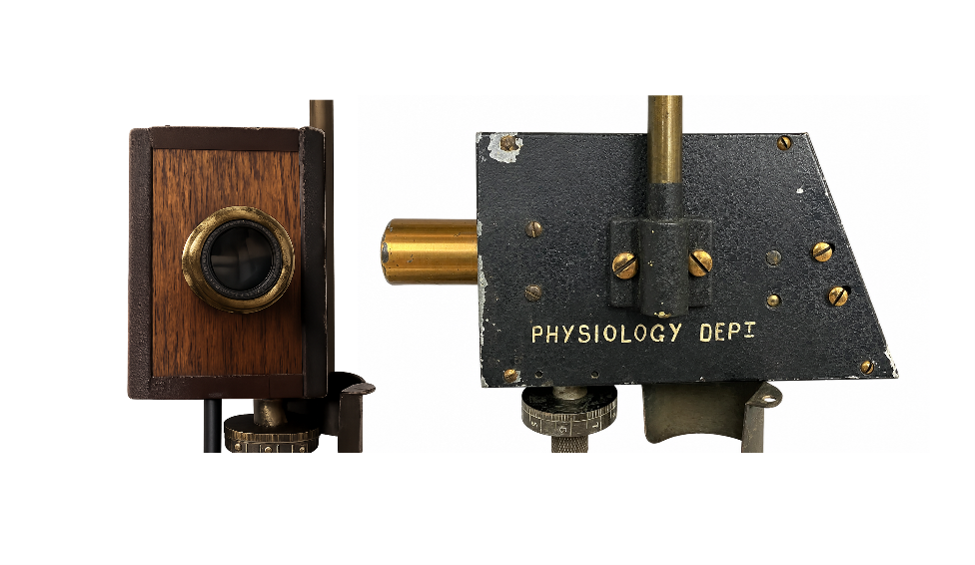
In the UCL Historic Objects and Collections series, the team explores the stories behind the physiological artefacts housed within the institute’s rich collection. The object featured in this blog is Physiological Instrument #019, a Hartridge reversion spectroscope. Developed in the early 1910s, this optical instrument was designed to precisely measure carbon monoxide levels in the blood. Its exceptional sensitivity made it an important tool in physiological research and contributed to the development of increasingly precise quantitative methods in physiology and biochemistry.
The silent killer: Carbon monoxide poisoning
In the early twentieth century, carbon monoxide poisoning was a major public health concern. The widespread use of coal gas, or “town gas,” for lighting, heating, and cooking, together with occupational exposure in industry, created a need to understand the causes of the gas’s remarkable toxicity. Because carbon monoxide is colourless and odourless, its detection and quantification posed a significant challenge for physicians. Early symptoms, including headache, dizziness, fatigue, and nausea, could easily be mistaken for common illnesses, while higher levels of exposure could rapidly lead to unconsciousness and death with little warning.
Carbon monoxide haemoglobin interaction
Researchers, most notably Claude Bernard and Felix Hoppe-Seyler, had shown that carbon monoxide binds to haemoglobin, preventing the normal transport of oxygen in the blood and providing the first clear explanation for the gas’s toxicity. However, accurately measuring the amount of carbon monoxide present in the blood remained a considerable challenge. Today, this can be achieved using photoelectric spectrophotometry, in which electronic detectors measure subtle changes in the absorption spectrum of haemoglobin caused by the formation of carboxyhaemoglobin. In the early twentieth century, however, such technology did not exist.
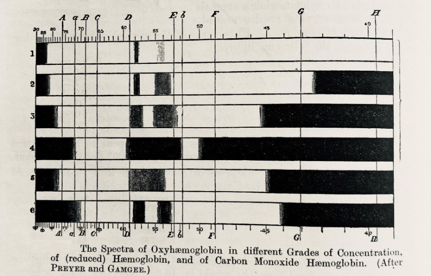
Instead, spectroscopic analysis relied on optical instruments that enabled observers to examine the absorption bands produced when light passed through a specimen. A prism separated white light into a spectrum of colours, much like a rainbow. As the light passed through a sample, specific wavelengths were absorbed, producing characteristic dark bands within the spectrum (Figure 2). These bands acted as optical fingerprints, allowing different forms of haemoglobin to be identified (1). However, the spectra of haemoglobin and carboxyhaemoglobin overlap extensively and differ only by a slight shift in the position of their absorption bands, making it extremely difficult to precisely measure carbon monoxide concentrations in the blood.
To overcome this problem, Hamilton Hartridge, working in the Physiological Laboratory at Cambridge, developed an innovative instrument known as the reversion spectroscope (2). Combining sophisticated optics with ingenious mechanical design, the instrument enabled exceptionally precise comparisons between spectral lines, allowing wavelength differences to be detected. This remarkable sensitivity made the reversion spectroscope particularly valuable for the quantitative measurement of carboxyhaemoglobin and helped establish new standards of precision in physiological research.
How does the Hartridge reversion spectroscope work?
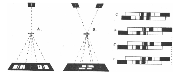
The Hartridge reversion spectroscope was designed to detect extremely small differences between absorption spectra. Light first passed through a specimen and then entered the instrument through a narrow slit, where it was collimated and dispersed by a prism into a spectrum of colours. A clever optical arrangement split the light into two spectra, one displayed in its normal orientation and the other reversed. Viewed through a single eyepiece, these appeared as two parallel horizontal spectra stacked one above the other (Figure 3).
Absorption bands produced by substances such as haemoglobin and carboxyhaemoglobin appeared as dark lines in both spectra. A precision micrometre screw allowed one spectrum to be shifted relative to the other; because one spectrum was reversed, the bands moved in opposite directions. By aligning corresponding spectral lines and measuring the displacement required to achieve coincidence, Hartridge could determine very small wavelength differences with exceptional accuracy (accurate to one ångström unit), making the instrument particularly valuable for the quantitative measurement of carbon monoxide in blood.
Hartridge published this work in 1912 in The Journal of Physiology (2) and later modified the method to calibrate the instrument (3). The reversion spectroscope was subsequently made available to a wider scientific community through commercial production by the Cambridge instrument maker W. H. Grayling and later by R. and J. Beck Ltd of London. In 1937, the instrument retailed for £25 and 4 shillings (approximately £2,000 in today’s currency) (4). The instrument preserved in the UCL collection was manufactured by W. H. Grayling.
Physiologist and inventor Hamilton Hartridge (1886-1976)
Hartridge studied at the University of Cambridge and graduated from St George’s Hospital Medical School, London, in 1914. He subsequently became a Fellow of King’s College, Cambridge, and Lecturer in the Physiology of the Special Senses at the University of Cambridge. Hartridge was renowned for his ingenuity in both experimentation and instrument design, particularly in the field of optics. His exceptional technical skills led to the development of several influential instruments and methodologies, including the reversion spectroscope and the continuous-flow apparatus for measuring the rates of extremely rapid chemical reactions.
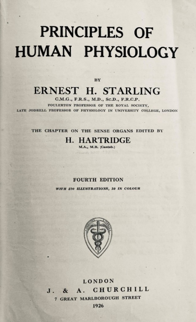
From 1927 to 1947, Hartridge served as Professor of Physiology at St Bartholomew’s Hospital Medical College, London (now part of Queen Mary University of London). He later became Director of the Vision Research Unit of the Medical Research Council, a position he held from 1947 to 1951. His appointment was announced in Nature, reflecting his standing within the scientific community (5). In addition to making important contributions to several areas of physiology, Hartridge contributed to Ernest Starling’s influential textbook Principles of Human Physiology by authoring the chapters on the physiology of the sense organs (Figure 4).
Hartridge’s scientific interests extended well beyond vision and spectroscopy. In 1920, he proposed that bats navigate and avoid obstacles by emitting sounds at high frequencies above the range of human hearing (6). Although the mechanism had not yet been experimentally demonstrated, this proved to be a remarkably accurate prediction of what is now known as echolocation.
Beyond carbon monoxide poisoning: Testing a fundamental question in physiology
Hartridge’s interest in carbon monoxide was not originally motivated by poisoning studies alone. Carbon monoxide provided a powerful experimental tool for studying pulmonary gas exchange because its binding to haemoglobin could be measured spectroscopically. At the beginning of the twentieth century, many physiologists accepted the possibility that the lung was not merely a passive membrane. John Scott Haldane argued that the pulmonary epithelium might actively secrete oxygen into the blood (7). In contrast, the Danish physiologists, August and Marie Krogh, developed a quantitative theory showing that oxygen transfer across the lung could be fully explained by passive diffusion (8). Through a series of elegant experiments and mathematical analyses, Marie Krogh demonstrated that the alveolar membrane possessed sufficient permeability and surface area for diffusion to account for oxygen uptake (9).
In another 1912 paper in The Journal of Physiology, Hartridge also addressed this fundamental question by using carbon monoxide as a tracer gas to investigate how gases pass from the lungs into the blood (10). By precisely measuring carbon monoxide uptake using spectroscopic methods, he found no convincing evidence for active oxygen secretion by the lungs. Instead, his results supported the emerging view that gas exchange occurs primarily by passive diffusion across the alveolar membrane, providing evidence consistent with the diffusion theory advanced by August and Marie Krogh.
Monitoring extremely rapid chemical reactions
Hartridge’s innovative reversion spectroscope not only advanced physiological research but also contributed significantly to biochemistry by enabling the study of extremely rapid chemical reactions. In 1927, Hartridge and Francis J. W. Roughton introduced the continuous-flow apparatus, a pioneering instrument that enabled the measurement of chemical reactions occurring within milliseconds (11). The ingenious principle behind the device was its conversion of time into distance: reacting solutions were rapidly mixed and passed through a narrow observation tube at a known speed, so that each position along the tube represented a precise interval of time after mixing. By examining changes in the haemoglobin spectrum at different points along the flow path, Hartridge and Roughton could determine how quickly oxygen, carbon monoxide, and other gases combined with haemoglobin.
This breakthrough laid the foundations of modern rapid-reaction kinetics and influenced the development of later stopped-flow techniques widely used in biochemistry and enzymology. The apparatus was closely linked to Hartridge’s earlier reversion spectroscope, extending its highly precise spectroscopic methods from measuring the extent of a reaction to measuring its rate.
Hartridge’s innovations influenced multiple scientific disciplines and demonstrate the importance of developing new instruments and experimental techniques in physiology. Despite its humble appearance, this wooden optical device occupies an important place in the history of science, contributing not only to the measurement of carbon monoxide in the blood but also to the testing of fundamental physiological theories and the development of quantitative methods that shaped modern physiology and biochemistry.
Acknowledgment: The authors are grateful to Liz Blanks and Ignacio Echeverria Faccin (UCL Museums & Cultural Collections) for their collaboration and expert advice.
If you missed the first five artefacts on display by the UCL Historic Objects and Collections team, read their blogs, Bárány’s Box, the Kymograph, Haldane apparatus, From Wills’ factor to folic acid and From squid giant axons to recording action potentials inside a nerve fibre to discover more about the history of physiology.
References
- Starling EH and Hartridge H. Principles of Human Physiology. 4th ed. London: J. & A. Churchill; 1926.
- Hartridge H. A spectroscopic method of estimating carbon monoxide. The Journal of Physiology. 1912;44(1-2):1-21. doi: 10.1113/jphysiol.1912.sp001496.
- Hartridge H. Calibration of the reversion spectroscope for the estimation of CO in blood. The Journal of Physiology. 1922;57(1-2):47-51. doi: 10.1113/jphysiol.1922.sp002041.
- R. and J. Beck Ltd. The Hartridge reversion spectroscope. Journal of Scientific Instruments. 1937;14:385.
- Prof. Hamilton Hartridge, F.R.S. Nature. 1948;161(4080):49. doi:10.1038/161049a0.
- Hartridge H. The avoidance of objects by bats in their flight. The Journal of Physiology. 1920;54(1-2):54-7. doi: 10.1113/jphysiol.1920.sp001908.
- Douglas CG, Haldane JS. The causes of absorption of oxygen by the lungs. Journal of Physiology. 1912;44(4):305–354. doi:10.1113/jphysiol.1912.sp001518.
- Krogh A, Krogh M. On the rate of diffusion of carbonic oxide into the lungs of man. Skandinavisches Archiv für Physiologie. 1910;23:236–247. doi/10.1111/j.1748-1716.1910.tb00600.x
- Krogh M. The diffusion of gases through the lungs of man. The Journal of Physiology. 1915;49(4):271–300. doi:10.1113/jphysiol.1915.sp001710.
- Hartridge H. Experiments on the oxygen secretion in the lung of man by the carbon monoxide method. The Journal of Physiology. 1912;45(3):170-81.
- Hartridge H, Roughton FJ. The rate of distribution of dissolved gases between the red blood corpuscle and its fluid environment: Part I. Preliminary experiments on the rate of uptake of oxygen and carbon monoxide by sheep’s corpuscles. The Journal of Physiology. 1927;62(3):232-42. doi: 10.1113/jphysiol.1927.sp002354.