

By Sadali Wanniarachchi (The University of Manchester) and Alireza Mani (UCL), UK
The Historic Objects and Collections at UCL series explores physiological artefacts preserved within the Institute’s collection. The object featured in this blog is Physiological Instrument #059, an Inductorium (also called an induction coil) that was widely used in 19th- and early-20th-century physiology laboratories to stimulate nerves and muscles with brief electrical pulses.
Early days of electrical stimulation
Coming off the first wave of evidence for the existence of animal electricity (Galvani, 1791), the field of 19th century physiology had become fascinated by the study of nerve stimulation. If neural inputs could be precisely manipulated and their physiological consequences mapped, the mechanisms underlying communication between tissues could be deciphered. However, physiologists sought robust and reliable techniques that would enable them to stimulate excitable tissues with greater precision and accuracy.
Yet for several decades following its emergence as a field, electrophysiology was confined to the mere observational – closed off to standardisation by the methods of the time, which could not be reliably regulated nor evoke sustained muscle contraction. In 1846, that constraint was lifted by the development of the specialised inductorium (induction coil). Hinged on the principles of electromagnetism, the inductorium provided researchers with the means to systematise pulse delivery and, for the first time, reproducibly parse the relationship between stimulus input and output. As a result, scientists “…need no longer misunderstand each other by experimenting with apparatus which did not admit of comparison, but… now compare notes with the greatest accuracy” (Hearder, 1872). By liberating the field into the realm of quantification, the induction coil spearheaded advances across physiology, furnishing discoveries from bench to bedside.
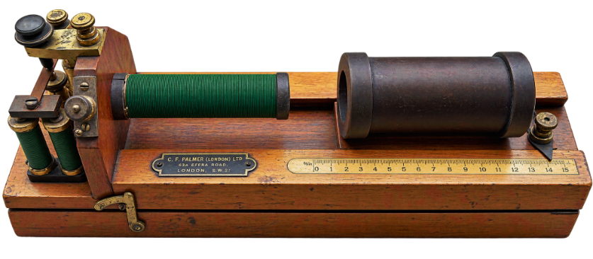
Figure 1. Inductorium manufactured by C. F. Palmer Ltd, on display as part of the UCL Physiology Collection (Object LDUSC-PHYSIO-59).
Development of the inductorium
The inductorium owes its birth to a collision of designs from across the globe. Comprised of a copper winding assembly and iron core, the earliest induction coil was developed by Irish priest Nicholas Callan and American scientist Charles Grafton Page, in 1836. Within a decade, German physiologist Emil Du Bois-Reymond (1818 –1896) had adapted their prototype to physiological research, improving it with the integration of modules like an adjustable secondary position, an automatic interrupter, and a short-circuiting key (Geddes, 1994) (Figure 2).
How does the inductorium work?
A primary coil was placed in or around an iron core and slid into a secondary coil, which was made of the same material but had a greater number of turns. A battery-driven current would flow through the primary coil, thus generating a magnetic field around the iron core. As the field built, a make shock would be induced in the secondary coil. This voltage would be larger than that of the primary due to the greater number of secondary turns. By modulating the position of the windings relative to each other along a sliding sledge, the intensity of the induced/Faradic current could be refined (i.e., the closer the distance, the greater the pulse).
Following the make shock, the battery would then be switched off, producing a sudden drop in current and demagnetising the core. As the magnetic field collapsed to nil almost instantly (as opposed to the more gradual rise induced prior), a pulse of even larger voltage would be produced, known as a break shock. The battery would then be switched on again and the cycle resumed. This succession of make and break shocks from the secondary winding would be dispensed via binding posts through to the tissue of the experimental subject.
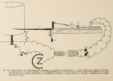
Figure 2. Illustration of the use of an inductorium (induction coil) to stimulate an isolated muscle and record its contraction in vitro using a kymograph. The figure is reproduced from Edward Schäfer’s 1912 textbook Experimental Physiology (Schäfer, 1912). The symbols C and Z indicate the copper (C) and zinc (Z) elements of the voltaic cell; a type of battery commonly used in physiological laboratories at the time. k′ indicates the short-circuiting key, or Du Bois-Reymond key, used to control the delivery of induced electrical stimuli to the preparation.
Further refinement of inductorium
The cycling of the pulse train could be further refined by the incorporation of an automatic interrupter/Neef’s hammer (Figure 3) and a short-circuiting key (K’ in Figure 2). Comprised of a ferromagnetic armature and spring, the automatic interrupter minimised the need for human intervention in stimulus delivery. During make as the iron core became magnetised by the flowing current, the armature would be summoned towards it and break the circuit. As a result, the magnetic field would collapse and demagnetise the core, reverting the armature to its original position and the series would be closed once again.
Later models like the Harvard inductorium also included a short-circuiting key to shunt only the break pulse to tissues. As the make shock resulted from a more gradual change in primary current, it produced a lower induced voltage in the secondary coil and was therefore less reliable in exceeding the threshold for nerve stimulation. To generate a more predictable and systematic sequence of stimuli, the secondary circuit could be short-circuited during make, allowing only the stronger and more reliable break shock to reach the preparation (Geddes et al., 1989).
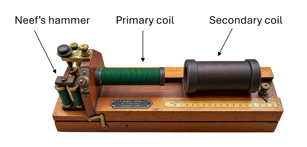
Figure 3. Palmer Ltd. inductorium (Du Bois-Reymond-type induction coil), c. 1930–1940, showing the Neef’s hammer, primary coil and sliding secondary coil. UCL Physiology Collection (Object LDUSC-PHYSIO-59)
Through these different manipulations of coil assembly, reliable nerve stimulation progressively became more attainable, skyrocketing the popularity of the device and its different variations. The specifications of a standard Palmer model, for example, are described in a 1932 paper by Ivan de Burgh Daly and Ulf von Euler, (1970 Nobel laurate): “…a Du Bois- Reymond induction coil (C. F. Palmer, Ltd.) is used for excitation. These Palmer coils have approximately 300 turns of copper wire in the primary, wound on an iron cone 5/8-inch diameter. The secondary coil consists of approximately 7500 turns of enamel copper wire amid the normal frequency of the vibrational ranges between 60 and 80 per second. Two dry cells giving 2-3 volts on load produce a secondary current which is just bearable on the tongue with the coils 9 cm apart.” (Daly and von Euler, 1932). The controlled electrical stimulation provided by the inductorium contributed to experiments that established fundamental principles of neurophysiology, including the localisation of motor function in the cerebral cortex and the excitatory and inhibitory effects of nerve stimulation (Ferrier, 1873; Geddes et al., 1989).
Inductorium in teaching and clinical practice
Naturally, the inductorium soon became part of the key infrastructure of teaching labs, with prominent 19th century professors recommending that “An induction coil generating primary and secondary induced currents, similar to the ordinary medical induction apparatus, should be put in the hands of each student” (Herdman et al., 1895). As illustrated in the 1880 UCL physiology syllabus, written by John Burdon-Sanderson, the first Jodrell Professor of Physiology, the inductorium was used across a wide range of physiological teaching, from cardiac excitation to skeletal muscle contraction (Burdeon-Sanderson, 1880). Beyond the bench, the device moved the needle translationally too, pioneering much of electrotherapeutics as we know it today.
In the mid-nineteenth century, the French physician Guillaume Duchenne de Boulogne pioneered the use of induction currents for the localised electrical stimulation of nerves and muscles. His method, which he termed faradisation in honour of Michael Faraday, helped establish electrical stimulation as a tool for investigating neuromuscular function. He reports in a series of case studies that “…there is no other therapeutic agent the action of which is comparable to that of cutaneous faradisation…” (Duchenne, 1871). By providing the degree of control and reliability over stimulation that it did, the inductorium revealed to physicians the clinical value of neuromodulation, thus serving as the progenitor of contemporary stimulators used for treatment today.
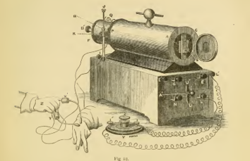
Figure 4. Illustration of an induction-coil apparatus used by Guillaume Duchenne for the localised electrical stimulation of nerves and muscles in clinical experiments (1870s). Surface electrodes connected to the apparatus are shown being applied to the hand and forearm. Reproduced from Duchenne’s A Treatise on Localized Electrization, and Its Applications to Pathology and Therapeutics (Duchenne, 1871)
The transition to electronic stimulation
Despite its nearly century-long reign in physiology, the specialised inductorium finally succumbed to the surge of industrial progress in the 1940s that accompanied World War II. The advent of solid state circuity revolutionised research and dethroned swathes of devices across medicine, from relay-based instruments and vacuum-tube electronics to the specialised inductorium (Cullen, 1941; Geddes, 1994). These highly integrated circuits, comprised of transistors, and later microprocessors, were not only more minimalist and practical than the induction coil, but also far superior in their honing of pulse parameters and output resolution (Geddes, 1994; Petraglia, 1946). By facilitating the progressively precise probing of increasingly subtle physiological processes, solid state electronics accelerated progress in research exponentially, and did so with an elegance that required neither mechanical interruption nor bulky magnetic apparatus.
While the inductorium of the 1800s has all but vanished from the lab, it survives in the ideas that it galvanised into action. By steering the field from the demonstrative to the experimental, the induction coil helped carve the ground of physiology into tractable paths. Now replaced with an electronic lineage that relentlessly becomes smaller, faster, and more efficient, it is worth remembering the device that was once “…one of the most familiar and useful instruments in the physiological laboratory…” (Martin, 1912). Beyond the display of the model of the inductorium at the UCL Historic Objects and Collections centre, its principles can still be recognised in many of the familiar and useful instruments of today.
Acknowledgment: The authors are grateful to Alanna Orpen (The Physiological Society) for her collaboration and expert advice.
Discover more about the history of physiology. If you’ve missed the previous blogs by the UCL Historic Objects and Collections team, you can read through the series from Bárány’s Box to the Kymograph to the Haldane apparatus. Enjoy more showcases of physiologists and artefacts with From Wills’ factor to folic acid; From squid giant axons to recording action potentials inside a nerve fibre; Measuring the invisible: The Hartridge Reversion Spectroscope; Winifred Cullis: Bringing human physiology to the public, From mentor to pupil: Starling, Verney, and the evolution of the heart-lung-kidney preparation and Measuring muscle heat: AV Hill and the Thermopile
References
Cullen, A.B. Jr (1941) ‘An A.C. operated electronic inductorium’, Science, 94(2435), pp. 219–220. doi: 10.1126/science.94.2435.219
Daly, I. de B. and von Euler, U.S. (1932) ‘The functional activity of the vasomotor nerves to the lungs in the dog’, Proceedings of the Royal Society of London. Series B, Containing Papers of a Biological Character, 110, pp. 92–111. doi: 10.1098/rspb.1932.0013.
Duchenne, G.-B. (1871) A treatise on localized electrization, and its applications to pathology and therapeutics. Translated from the 3rd French edn by H. Tibbits. Philadelphia, PA: Lindsay & Blakiston. doi: 10.5962/bhl.title.24567.
Ferrier, D. (1873) ‘Experimental researches in cerebral physiology and pathology’, Journal of Anatomy and Physiology, 8, pp. 152–155.
Galvani, L. (1791) De viribus electricitatis in motu musculari commentarius. Bononiae
Geddes, L.A. (1994) ‘The first stimulators: Reviewing the history of electrical stimulation and the devices crucial to its development’, IEEE Engineering in Medicine and Biology Magazine, 13(4), pp. 532–542. doi: 10.1109/51.310995.
Geddes, L.A., Foster, K.S., Senior, J.E. and Kuhfeld, A. (1989) ‘The inductorium: The stimulator associated with discovery’, Biomedical Instrumentation & Technology, 23(4), pp. 308–313.
Hearder, J.N. (1872) ‘On the progress of electro-therapeutics, with a description of galvanic instruments invented by the author’, Transactions of the Devonshire Association for the Advancement of Science, Literature and Art, pp. 107–116
Herdman, W.J. et al. (1895) An international system of electro-therapeutics for students, general practitioners, and specialists. Philadelphia, PA: F.A. Davis Company.
Martin, E.G. (1912) The measurement of induction shocks: A manual for the quantitative use of Faradic stimuli. New York: John Wiley & Sons.
Petraglia, F.A. (1946) The electronic engineering master index: A subject index to electronic engineering periodicals, January 1925 to June 1945. New York: Macmillan.
Burdon-Sanderson, J. (1880) Syllabus of a course of lectures on physiology. 2nd edn. Philadelphia, PA: Lindsay & Blakiston
S Schäfer, E.A. (1912) Experimental physiology. London: Longmans, Green and Co.